
Quick answer
What Is Noncardiogenic Pulmonary Edema?
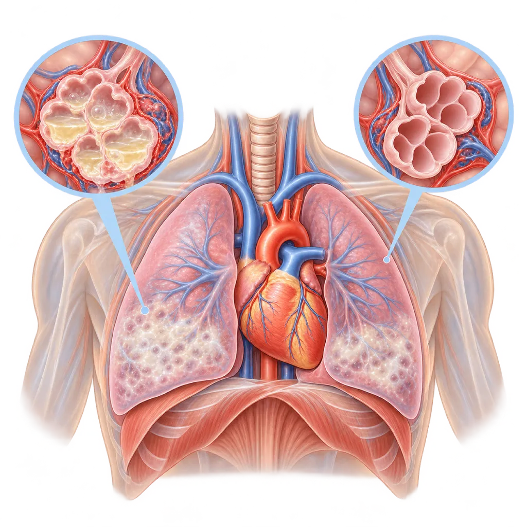
Noncardiogenic pulmonary edema is a buildup of fluid in the air sacs of the lungs that is not caused by the heart but by leaky lung blood vessels due to severe infection, injury, or other triggers. It is an emergency and needs treatment in a hospital.
Sources01 / Understanding the condition
About Noncardiogenic Pulmonary Edema
In healthy lungs, the air sacs (alveoli) have very thin, dry walls, so oxygen passes easily into the blood. Pulmonary edema means the air sacs and the tissue around them fill with fluid. In cardiogenic pulmonary edema, fluid is pushed out by high pressure from a weak heart or a faulty valve, covered on the Cardiogenic Pulmonary Edema page. In noncardiogenic pulmonary edema, the pressure from the heart is usually normal, but the walls of the lung blood vessels and air sacs are damaged and leaky, so protein-rich fluid floods the lungs.
Known Forms
The main form is acute respiratory distress syndrome (ARDS), which can be triggered by severe pneumonia including COVID-19, sepsis, inhaling stomach contents, severe injury, inflammation of the pancreas, or a blood transfusion. Other forms are less common: high-altitude pulmonary edema in climbers who ascend too quickly, neurogenic pulmonary edema after a head injury or bleeding in the brain, pulmonary edema caused by medicines or chemical gases, and edema after a collapsed lung is re-expanded. In ARDS, shortness of breath usually appears within a few hours to one week after the trigger, often while the patient is already in the hospital for another illness.
Why a Heart Cause Needs to Be Ruled Out
The two can look similar on a chest X-ray, yet they are treated differently. In edema caused by the heart, fluid-removing medicines (diuretics) and heart medicines relieve symptoms quickly. In noncardiogenic edema, the key to treatment is dealing with the trigger and supporting breathing until the lungs recover. In some patients both occur at the same time, so doctors assess the heart with echocardiography and blood tests. A normal heart size, a normal heart filling pressure, and a low NT-proBNP level support a noncardiogenic cause.
Care and Recovery
Most patients are treated in the intensive care unit. Treatment includes oxygen, breathing support with lung-protective settings, lying face down (prone positioning) in severe cases, and careful fluid management. Recovery can take weeks to months. After going home, some people still feel weak or short of breath, or have trouble concentrating, so gradual exercise and regular follow-up help them return to their activities.
02 / Understanding the condition
Symptoms to Recognize.
Common Symptoms
- Shortness of breath that worsens over a few hours to a few days
- Fast, shallow breathing
- Cough, sometimes with frothy sputum
- Feeling very tired or restless due to a lack of oxygen
- Fast heart rate
- Symptoms of the triggering illness, such as high fever and chills in a severe infection
Symptoms in Children
Children often cannot explain that they are short of breath. Watch how they breathe, especially during a fever, after choking, or after nearly drowning.
- Breathing much faster than usual
- The chest wall or the spaces between the ribs pulling inward with each breath
- Flaring nostrils
- Grunting sounds when breathing out
- Looking limp or fussy, or having trouble eating and drinking
03 / Understanding the condition
What Causes This Condition?
- Severe infection and sepsis, including severe pneumonia caused by bacteria or viruses such as influenza and COVID-19
- Inhaling stomach contents into the airways (aspiration) or nearly drowning
- Severe injury, extensive burns, or severe inflammation of the pancreas
- A lung reaction to a blood transfusion, called transfusion-related acute lung injury
- Inhaling smoke from a fire or chemical gases that damage the airways
- Ascending too quickly to altitudes above about 2,500 meters
- Head injury, bleeding in the brain, or severe seizures that trigger neurogenic pulmonary edema
- A reaction to certain medicines or substances, including an opioid overdose
04 / Understanding the condition
Who Is at Higher Risk?
Medical
Sepsis, severe pneumonia, severe injury, inflammation of the pancreas, major surgery, and repeated blood transfusions. Chronic lung disease and weak immunity can make the condition worse.
Lifestyle
Long-term excessive alcohol use, smoking, and drug misuse, including opioids, are linked to a higher risk of acute lung injury.
Age
In older age, recovery is slower and severe illness is more common. In children, respiratory infections, choking, and drowning are triggers to watch for.
Other
Ascending quickly to high altitude, a previous episode of high-altitude pulmonary edema, diving, and exposure to smoke or chemical gases at work.
Groups at Higher Risk
- Patients with Severe Infection or Sepsis: Inflammation that spreads throughout the body can damage the lung blood vessels. Shortness of breath that increases during a high fever needs to be checked right away.
- Patients with Severe Injuries and Burns: Extensive injuries, burns, and large-volume blood transfusions can trigger leaky lung blood vessels within the first few days.
- Climbers and Travelers to High Altitudes: Ascending too quickly to altitudes above about 2,500 meters can trigger pulmonary edema, especially in people who have had it before.
- Children with Lung Infections or a History of Choking: Severe pneumonia, choking, and nearly drowning can trigger ARDS in children, which is managed with child-specific guidelines.
05 / Understanding the condition
How Is It Diagnosed?
The doctor first measures oxygen levels with a finger clip device and a blood gas analysis, then gives oxygen while looking for the cause. A chest X-ray shows shadows of fluid in both lungs. To make sure the fluid does not mainly come from the heart, the doctor performs echocardiography to assess the heart's pumping, valves, and filling pressure, along with an NT-proBNP test; a low level makes a heart cause less likely.
According to the 2024 global definition of ARDS, ARDS is diagnosed when shortness of breath appears within one week of a known trigger, there are shadows in both lungs that are not fully explained by fluid in the chest cavity, a collapsed lung, or heart failure, and blood oxygen levels fall by a defined measure. Bedside lung ultrasound can now also be used. A chest CT scan helps show the extent of the damage and its trigger, while blood tests and cultures look for infection and assess the function of other organs.
Chest X-Ray
Shows fluid shadows in both lungs. A normal heart size and the pattern of the shadows help distinguish it from edema caused by the heart.
Blood gas analysis and oximetry: measure oxygen and carbon dioxide levels to assess severity and monitor breathing support.
Echocardiography
Assesses the heart's pumping strength, valves, and filling pressure to make sure the fluid in the lungs does not mainly come from the heart.
NT-proBNP and BNP Test
A low level makes a heart cause less likely, although the result is still read together with other tests.
Chest CT scan: shows the spread of fluid and lung damage in more detail, and looks for triggers such as pneumonia or aspiration.
Bedside lung ultrasound: a quick, radiation-free test to look for signs of fluid in the lungs in patients who are hard to move.
Laboratory Tests
Complete blood count, infection markers, blood cultures, kidney function, and liver function to look for triggers and assess other organs.
06 / Understanding the condition
Treatment Options.
Treatment is decided by a doctor after an assessment. The categories below can help you follow the conversation with your doctor.
Treating the Trigger
Antibiotics for infection, stopping the transfusion or the triggering medicine, and descending from altitude with oxygen for high-altitude pulmonary edema. These steps are the foundation of recovery.
Oxygen and Breathing Support Without a Tube
Oxygen through a mask or a high-flow nasal cannula, or a pressurized mask, to raise oxygen levels without inserting a breathing tube.
Lung-Protective Ventilation
If breathing support without a tube is not enough, the ventilator is set with small breath volumes and limited pressure so the fragile lungs are not damaged further.
Prone Positioning
In moderate to severe ARDS, the patient is placed face down for many hours each day so air spreads more evenly and oxygen levels improve.
Fluid Management
Once circulation is stable, fluids are limited and fluid-removing medicines (diuretics) may be used so the lungs do not become wetter.
Corticosteroids in Selected Cases
The ATS 2024 guideline suggests corticosteroids for some patients with ARDS. The doctor decides after weighing the trigger and the risks.
Extracorporeal Membrane Oxygenation (ECMO)
A machine that temporarily takes over oxygen exchange, for a small number of patients in very severe condition, at experienced centers.
07 / Understanding the condition
Complications to Watch For.
Respiratory Failure
The lungs cannot meet the body's oxygen needs, so the patient needs a ventilator for a time.
Multiple Organ Dysfunction
A lack of oxygen and inflammation can affect the kidneys, liver, brain, and circulation. Monitoring in the intensive care unit helps detect this early.
Strain on the Right Side of the Heart
Lung damage and pressure from breathing support can raise the pressure in the lung blood vessels, putting strain on the right ventricle. Echocardiography is used to monitor this.
Additional Infections During Hospital Care
Long-term use of a ventilator and catheters increases the risk of pneumonia and bloodstream infections.
Air Leak from the Lung
Fragile lungs can leak, so air collects in the chest cavity (pneumothorax) and needs to be drained with a tube.
Lung Scarring
In some patients, the lungs heal with scar tissue that makes shortness of breath during activity last longer.
Muscle Weakness and Problems After Intensive Care
After a long stay in intensive care, some people have muscle weakness, memory problems, anxiety, or sadness that improve gradually with rehabilitation.
08 / Understanding the condition
What Can You Do?
- Get checked right away if shortness of breath increases during a fever or lung infection, so treatment can start early.
- When climbing, ascend gradually; above 3,000 meters, raise your sleeping altitude by about 500 meters per day.
- Descend from altitude if shortness of breath and cough appear and do not improve with rest.
- Avoid inhaling smoke from fires or chemical gases, and use respiratory protection at work.
- Use medicines as prescribed and keep opioid medicines out of the reach of others.
- Quit smoking and limit alcohol to keep your lungs resilient.
- Stay up to date with influenza, pneumococcal, and COVID-19 vaccines as recommended by your doctor.
- Supervise children while they eat and when they are near water to prevent choking and drowning.
09 / Understanding the condition
When Should You See a Doctor?
Noncardiogenic pulmonary edema usually develops over hours to days and needs treatment in a hospital. Go to the emergency room right away or call emergency services if shortness of breath gets worse, especially during a severe infection, after an injury, choking, nearly drowning, inhaling smoke, or receiving a blood transfusion, or while at high altitude.
- Shortness of breath that worsens within a few hours
- Fast breathing with bluish lips or nails
- Confusion or extreme sleepiness while short of breath
- Shortness of breath and cough at high altitude that do not improve with rest
- In children: fast breathing, the chest pulling inward, or grunting
- After coming home from intensive care: shortness of breath, weakness, or trouble concentrating that does not improve; mention it at your follow-up visit
10 / Common questions
Questions You May Have.
Frequently asked questions about noncardiogenic pulmonary edema.
How is noncardiogenic pulmonary edema different from pulmonary edema caused by the heart?
Both fill the lungs with fluid, but the source is different. In edema caused by the heart, fluid is pushed out by high pressure from a weak heart or a faulty valve. In noncardiogenic edema, the heart pressure is usually normal, but the lung blood vessels are damaged and leaky. This difference determines treatment, so the doctor checks the heart with echocardiography and blood tests. Edema caused by the heart is covered on the Cardiogenic Pulmonary Edema page.
Is ARDS the same as noncardiogenic pulmonary edema?
ARDS is the main form of noncardiogenic pulmonary edema. The term ARDS is used when leaking of the lung blood vessels occurs within one week of a known trigger, such as pneumonia or sepsis, causes shadows in both lungs, and lowers oxygen levels by a defined measure. Noncardiogenic pulmonary edema also includes other forms, such as high-altitude pulmonary edema or edema after a head injury, which are partly treated differently.
Why are patients with ARDS placed face down?
When lying on the back, fluid and the weight of the heart press on the back part of the lungs, even though that part has many blood vessels. Lying face down spreads air more evenly, so oxygen levels often improve. The ESICM 2023 guideline recommends this position for many hours each day in moderate to severe ARDS on a ventilator. The intensive care team positions the patient carefully and protects the face, eyes, and skin while the patient is face down.
Can the lungs recover after ARDS?
Many patients recover and return to their activities, although recovery can take months. Lung function often improves gradually over six to twelve months. Some people still get short of breath during activity, tire easily, have muscle weakness, or have memory and mood problems after intensive care. Rehabilitation, gradual exercise, and regular follow-up help recovery. Mention any lasting symptoms to your doctor at your follow-up visits.
How can I prevent pulmonary edema when climbing mountains?
Ascend gradually. Above about 3,000 meters, the Wilderness Medical Society 2019 guideline recommends that your sleeping altitude increase by no more than about 500 meters per day, with a rest day every three to four days. If you become short of breath at rest, develop a cough, or your walking ability drops sharply, descend right away and seek help. People who have had high-altitude pulmonary edema should consult a doctor before climbing again.
Can noncardiogenic pulmonary edema happen again?
Yes, depending on the trigger. High-altitude pulmonary edema tends to recur in people who have had it before if they ascend at the same speed, so a slower climbing plan is needed. ARDS can occur again if a severe infection or another trigger arises. Vaccination, early treatment of infections, quitting smoking, and avoiding exposure to smoke or chemical gases help lower this risk.
Sources and Medical Review
Translated from the Indonesian edition medically reviewed by dr. Santi R. Dewayanti, Sp.P.
Sources as cited in the Indonesian edition.
- Matthay MA, Arabi Y, Arroliga AC, Bernard G, Bersten AD, Brochard LJ, et al. A New Global Definition of Acute Respiratory Distress Syndrome. Am J Respir Crit Care Med. 2024;209(1):37-47.
- Qadir N, Sahetya S, Munshi L, Summers C, Abrams D, Beitler J, et al. An Update on Management of Adult Patients with Acute Respiratory Distress Syndrome: An Official American Thoracic Society Clinical Practice Guideline. Am J Respir Crit Care Med. 2024;209(1):24-36.
- Grasselli G, Calfee CS, Camporota L, Poole D, Amato MBP, Antonelli M, et al. ESICM guidelines on acute respiratory distress syndrome: definition, phenotyping and respiratory support strategies. Intensive Care Med. 2023;49(7):727-59.
- Bos LDJ, Ware LB. Acute respiratory distress syndrome: causes, pathophysiology, and phenotypes. Lancet. 2022;400(10358):1145-56.
- Emeriaud G, López-Fernández YM, Iyer NP, Bembea MM, Agulnik A, Barbaro RP, et al. Executive Summary of the Second International Guidelines for the Diagnosis and Management of Pediatric Acute Respiratory Distress Syndrome (PALICC-2). Pediatr Crit Care Med. 2023;24(2):143-68.
- Luks AM, Auerbach PS, Freer L, Grissom CK, Keyes LE, McIntosh SE, et al. Wilderness Medical Society Clinical Practice Guidelines for the Prevention and Treatment of Acute Altitude Illness: 2019 Update. Wilderness Environ Med. 2019;30(4S):S3-18.
- Vlaar APJ, Toy P, Fung M, Looney MR, Juffermans NP, Bux J, et al. A consensus redefinition of transfusion-related acute lung injury. Transfusion. 2019;59(7):2465-76.
Doctors Who Treat This Condition

dr. Santi R. Dewayanti, Sp.P
Pulmonology
Clinical focusLung and Respiratory Disease · Asthma and COPD
View Profile
dr. Siska S. Danny, Sp.JP(K)
Interventional Cardiology
Coronary Intervention Procedures
Clinical focusPCI for Acute Coronary Syndrome · Primary Coronary Intervention
View Profile
dr. Tartila, Sp.A, Subsp.ETIA(K)
Pediatric Emergency and Intensive Care
Congenital and Structural Heart Center
Clinical focusPediatric Intensive Care · Pediatric Emergency Care
View Profile