
Quick answer
What Is Echocardiography?
Echocardiography, or a heart echo, is a heart ultrasound that uses sound waves to show heart chamber size, wall thickness, valve movement, and pumping strength. It is used to assess shortness of breath, swelling, or a heart murmur, and to monitor heart disease. An echo through the chest takes about 30 to 60 minutes, without radiation.
Sources01 / Understanding the procedure
About Echocardiography
What Is Echocardiography
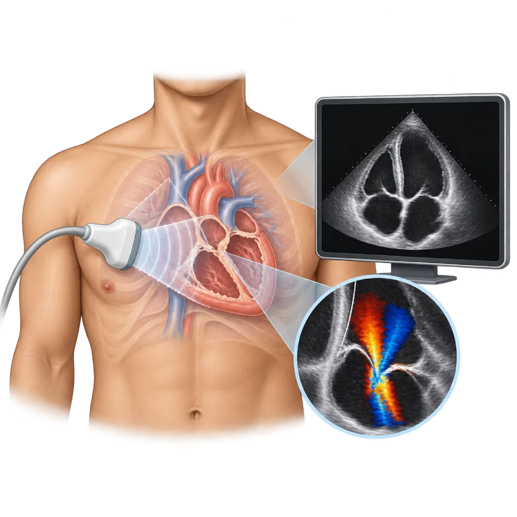
Echocardiography, often called a heart echo, is an ultrasound test of the heart. A small device called a probe sends out high-frequency sound waves that cannot be heard, then picks up the echoes that bounce back from the heart. A computer turns these echoes into moving images of the heart, so the doctor can watch your heart beating live on the screen. This test does not use radiation and can be repeated if needed.
What Is Assessed
An echo shows the size of all four heart chambers, the thickness and movement of their walls, and the shape and movement of the valves. From these images, the doctor calculates the ejection fraction, which is the percentage of blood the left ventricle pumps out with each beat and the main measure of the heart's pumping strength. An echo can also show fluid in the sac around the heart, clots inside the heart chambers, and holes in the wall that divides the heart chambers.
Doppler for Blood Flow
The Doppler part of the echo assesses the direction and speed of blood flow, which is displayed in color and as graphs. With Doppler, the doctor can see valves that are narrowed or leaking, estimate the pressure inside the heart and in the blood vessels of the lungs, and assess how the heart fills with blood as it relaxes. The whooshing sound you may sometimes hear during the test is the sound of blood flow from the Doppler.
Transthoracic and Transesophageal Echo
The majority of echoes are done through the chest, called transthoracic echocardiography or TTE. When clearer images are needed, for example to look for infection or clots on a valve, to look for a clot in the left atrium before a heart rhythm procedure, or to plan a valve repair, the doctor may perform transesophageal echocardiography or TEE. In TEE, a small probe is passed through the mouth into the esophagus (food pipe), which lies directly behind the heart, with the help of a sedative.
Similar Tests
An echo can also be done while the heart is stressed with a treadmill or medication to look for narrowing of the coronary arteries, which is covered on the Stress Echocardiography page. If the echo images do not fully answer the doctor's questions, other imaging tests may be considered, such as a cardiac CT scan, which is covered on the Cardiac CT Scan page.
What the Results Mean
The echo result is a report on the size of the heart chambers, the ejection fraction, the condition of the valves, and the estimated pressures inside the heart. The doctor relates these results to your symptoms, physical examination, and ECG. An echo is often repeated periodically to monitor valve disease, heart failure, or the effects of certain medications on the heart, so changes can be detected before they cause symptoms.
02 / Understanding the procedure
When Is This Procedure Considered?
A doctor may consider this procedure in the situations below. The decision is always made after a thorough assessment.
- Shortness of Breath or Leg SwellingLooks for a cause in the heart, such as heart failure or valve disease.
- Heart MurmurAn extra sound heard when listening to the heart may come from a narrowed or leaking valve.
- Heart Failure and CardiomyopathyMeasures pumping strength and heart chamber size, and monitors the results of treatment.
- After a Heart AttackAssesses parts of the heart wall that have weakened and looks for complications.
- Congenital Heart DiseaseShows holes in the wall that divides the heart chambers or structural heart abnormalities present from birth.
- Suspected Valve Infection or Source of ClotsLooks for infection on a valve or clots inside the heart, often with TEE.
Situations to Consider First
- No Absolute Contraindication for TTEAn echo through the chest can be done in almost everyone, including during pregnancy.
- Severe Esophageal Disease for TEENarrowing, a tear, or surgery of the esophagus means TEE needs to be reconsidered.
- Active Gastrointestinal BleedingTEE is postponed or replaced with another test until the bleeding is controlled.
- Unstable Breathing or Blood PressureGiving a sedative for TEE is reconsidered until your condition improves.
03 / Before the procedure
How Should You Prepare?
An echo through the chest requires almost no special preparation. Preparation for TEE is different and will be explained by the team when your test is scheduled.
Food and medications. For an echo through the chest, you may eat, drink, and take your medications as usual.
Clothing. Wear a top that is easy to remove, because your chest needs to be uncovered during the test. You will be given a cover sheet or an examination gown.
If you are having TEE. Do not eat or drink for about 4 to 6 hours beforehand. Let the team know if you have trouble swallowing, an esophageal condition, or dentures. Arrange for someone to take you home, because you must not drive for the rest of that day after receiving a sedative.
Bring your records. Bring a list of your medications and any previous echo or ECG results so the doctor can compare them.
Unsure about the preparation instructions? Ask our team before the procedure.
04 / During the procedure
How Is the Procedure Done?
An echo through the chest is done in an echo examination room without anesthesia and usually takes about 30 to 60 minutes. TEE requires a numbing spray in the throat and a sedative, with the examination itself taking about 15 to 30 minutes plus monitoring afterward.
Lying Down and Attaching Electrodes
You lie down, usually turned onto your left side so the heart is closer to the chest wall. A few ECG electrodes are attached to your chest to record your heart rhythm during the recording.
Recording the Images
A staff member applies gel to your chest, then moves the probe to several spots, for example beside the breastbone, below the left nipple, and below the breastbone. You may be asked to hold your breath briefly or change position. The probe is pressed slightly so the images are clear, but it is not painful.
Doppler and Measurements
Doppler is switched on to assess blood flow through the valves and heart chambers. If the images are unclear, a special ultrasound contrast agent may be injected through an IV to make the borders of the heart walls clearer.
If TEE Is Performed
Your throat is sprayed with a numbing medicine and a sedative is given through an IV. A thin probe is passed through your mouth into the esophagus as you swallow. Your breathing, blood pressure, and oxygen level are monitored throughout the examination.
05 / Benefits and risks
Benefits and Risks Should Be Discussed Together.
Every medical procedure has benefits and risks. Your doctor will explain both for your situation before the procedure.
Benefits You Can Expect
- No RadiationUses sound waves, so it is safe to repeat, including during pregnancy.
- Seeing the Heart in MotionWall and valve movement appear live on the screen as the heart beats.
- Assesses Many Things at OnceChamber size, pumping strength, valves, and heart pressures in a single test.
- PainlessAn echo through the chest uses only gel and a probe placed on the skin of the chest.
- Easy to Repeat for MonitoringChanges in valve disease or heart failure can be followed over time.
The benefits of a procedure are not a promise of a cure. Ask how the results will affect your care plan.
Risks You Should Know
- Discomfort from the ProbePressure from the probe on the chest can feel slightly uncomfortable, but it is not harmful.
- Unclear ImagesChest shape, body weight, or lung disease can make images difficult to obtain.
- Reaction to Ultrasound ContrastRare and usually mild, such as a headache or a warm feeling.
- Sore Throat After TEEUsually mild and goes away within one to two days.
- Effects of the Sedative in TEESlowed breathing or a drop in blood pressure; monitored and managed by the team.
- Esophageal Injury in TEEVery rare; the doctor assesses the risk before the examination is done.
Risks can differ from person to person. A full explanation and consent are given after an individual assessment.
06 / After the procedure
What Is Recovery Like?
After an echo through the chest, the gel is wiped off and you can go home right away and resume your usual activities.
Test results. A cardiologist reviews the recording and writes a report. The results are discussed with you during your consultation, including whether further tests are needed.
After TEE. You are monitored until the effects of the sedative wear off, usually about 1 hour. Do not eat or drink until the numbness in your throat has gone. Your throat may feel slightly sore for one to two days.
Repeat echo. In valve disease, heart failure, or certain treatments, the echo is repeated on the schedule set by your doctor to monitor changes.
Warning signs to watch for after TEE. Contact the medical team right away or go to the emergency room if you experience:
- Pain when swallowing that gets worse or does not improve
- Vomiting blood or black stools
- Chest pain or shortness of breath
- Fever
Know the Signs That Need Prompt Care.
Follow the team's guidance on warning signs. If severe or sudden symptoms appear after the procedure, go to the nearest emergency department right away or call Heart Attack Center 1500 258.
07 / Team and support
Doctors and Procedure Support
The doctors below are listed for this procedure. Listed by name, not by ranking.

dr. BRM Ario S. Kuncoro, Sp.JP(K)
Echocardiography & Cardiovascular Imaging
Cardiovascular Diagnostic Center +2 centers
Clinical focusAdvanced and 3D Echocardiography · Transesophageal Echocardiography
View Profile
Dr. med. dr. Denio A. Ridjab, Sp.JP(K)
Interventional Cardiology
Coronary Intervention Procedures +2 centers
Clinical focusInterventional Cardiology · Coronary Artery Disease
View Profile
dr. Radityo Prakoso, Sp.JP(K)
Pediatrics & Congenital Heart Disease
Congenital and Structural Heart Center
Clinical focusPediatric Cardiology Consultation · Congenital Heart Disease Care
View Profile4D Echocardiography
An ultrasound scan of the heart that records three-dimensional images in real time to assess pumping function, the valves, and…
The availability of technology and test support is confirmed with the team. Technology is chosen according to clinical need.
08 / Common questions
Before You Plan the Procedure.
Frequently asked questions about echocardiography, from preparation to what the results mean.
Does a heart echo hurt?
An echo through the chest is not painful. You only feel the cool gel and light pressure from the probe on your chest, which can sometimes feel slightly uncomfortable around the ribs. No needles are used, unless the doctor needs to give an ultrasound contrast agent through an IV. In TEE, your throat is numbed and you are given a sedative, so it usually only feels uncomfortable when the probe is swallowed.
Do I need to fast before an echo?
For an echo through the chest, you do not need to fast and may take your medications as usual. Fasting is only needed if you are having TEE, usually for about 4 to 6 hours beforehand, so your stomach is empty when the probe is inserted and the sedative is given. The team will give you written instructions for your type of test.
How is an echo different from an ECG?
An ECG records the heart's electrical activity through electrodes attached to the chest, so it shows the rhythm and signs of disturbed blood flow. An echo shows images of the heart: chamber size, wall movement, valves, and pumping strength. The two complement each other. Doctors often request both, because a normal ECG does not necessarily mean the heart valves and pumping function are also normal.
When do I need TEE?
TEE is used when an echo through the chest has not given clear enough images. Examples include looking for infection on a valve, looking for a clot in the left atrium before a heart rhythm procedure, assessing an artificial valve, or planning a valve repair. Because the probe sits in the esophagus directly behind the heart, the valves and the wall between the chambers are seen more clearly. The doctor explains the reasons and risks before the examination.
How is echocardiography different from stress echocardiography?
Standard echocardiography is done while you lie at rest and assesses the structure and function of the heart. Stress echocardiography records images of the heart before and after the heart is stressed with a treadmill or medication, to see parts of the wall that weaken from a lack of blood when working hard. The test chosen depends on the doctor's question about your symptoms.
How often does an echo need to be repeated?
It depends on your condition. In mild valve disease, an echo may be repeated every few years, while in moderate to severe valve disease or heart failure, the intervals are shorter, as advised by your doctor. An echo is also repeated if your symptoms change, after a new treatment is started, or during certain cancer treatments that can affect the heart.
Sources and Medical Review
Translated from the Indonesian edition medically reviewed by dr. BRM Ario S. Kuncoro, Sp.JP(K).
Sources as cited in the Indonesian edition.
- Mitchell C, Rahko PS, Blauwet LA, et al. Guidelines for Performing a Comprehensive Transthoracic Echocardiographic Examination in Adults: Recommendations from the American Society of Echocardiography. J Am Soc Echocardiogr. 2019;32(1):1-64.
- Lang RM, Badano LP, Mor-Avi V, et al. Recommendations for Cardiac Chamber Quantification by Echocardiography in Adults: An Update from the American Society of Echocardiography and the European Association of Cardiovascular Imaging. J Am Soc Echocardiogr. 2015;28(1):1-39.e14.
- Hahn RT, Abraham T, Adams MS, et al. Guidelines for Performing a Comprehensive Transesophageal Echocardiographic Examination: Recommendations from the American Society of Echocardiography and the Society of Cardiovascular Anesthesiologists. J Am Soc Echocardiogr. 2013;26(9):921-964.
- McDonagh TA, Metra M, Adamo M, et al. 2021 ESC Guidelines for the diagnosis and treatment of acute and chronic heart failure. Eur Heart J. 2021;42(36):3599-3726.
- Praz F, Borger MA, Lanz J, et al. 2025 ESC/EACTS Guidelines for the management of valvular heart disease. Eur Heart J. 2025;46(44):4635-4736.
- Delgado V, Ajmone Marsan N, de Waha S, et al. 2023 ESC Guidelines for the management of endocarditis. Eur Heart J. 2023;44(39):3948-4042.