
Quick answer
What Is Arterial Embolism?
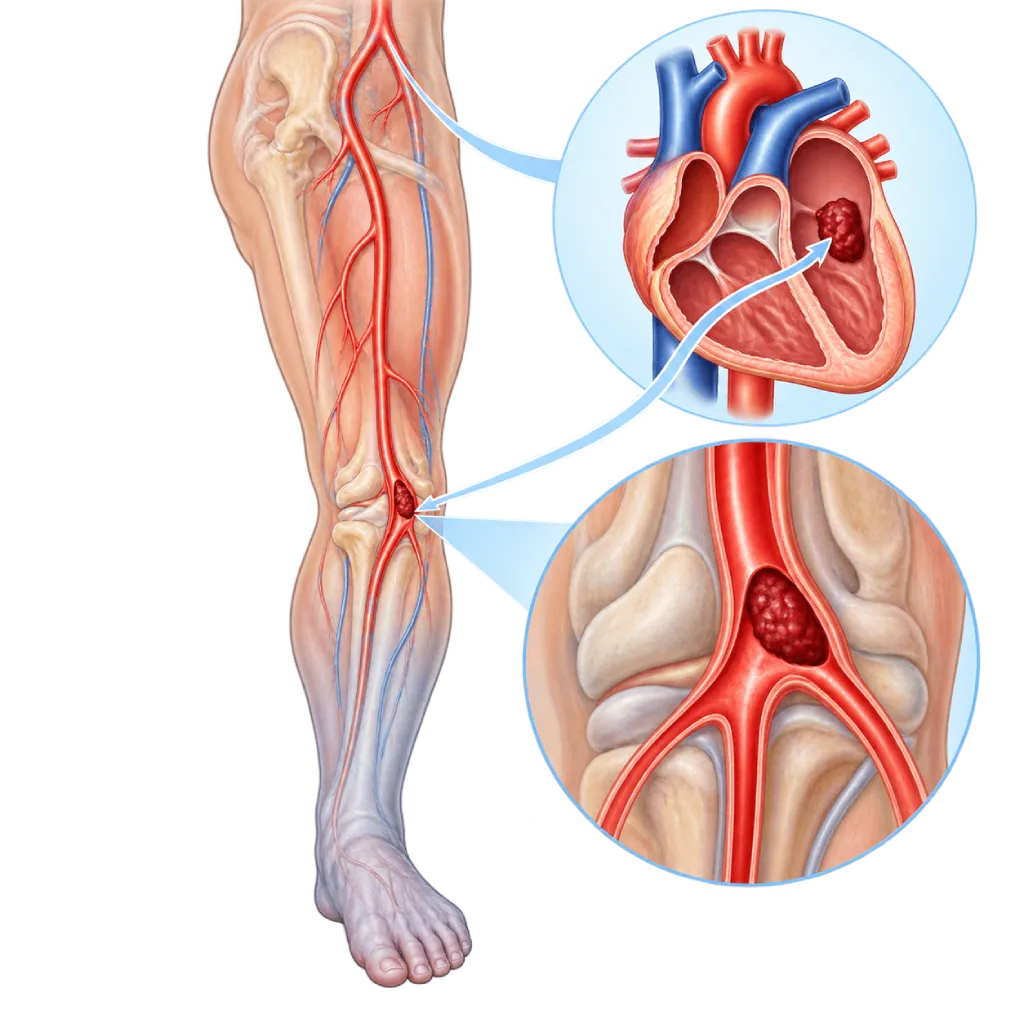
Arterial embolism is a sudden blockage of an artery by a blood clot or other material carried in the bloodstream, usually from the heart. The blocked leg or organ is starved of blood within minutes, so it needs urgent treatment in the emergency room.
Sources01 / Understanding the condition
About Arterial Embolism
Arterial embolism is a sudden blockage of an artery by material carried in the bloodstream from elsewhere, called an embolus. An embolus is usually a blood clot, but it can also be a fragment of plaque, a clump of bacteria from an infected heart valve, or part of a heart tumor. The embolus travels with the blood until it lodges in a smaller artery, instantly stopping blood flow to the tissue beyond it.
Where the Clot Comes From
In most cases, the embolus comes from the heart. A common cause is atrial fibrillation, a rhythm disorder that lets blood pool in the left atrium so it clots easily, covered on the Atrial Fibrillation page. Clots can also form in the left ventricle after a heart attack, on an artificial heart valve, or with a valve infection, covered on the Infective Endocarditis page. Other sources are a widened section of the aorta or of the artery behind the knee, and plaque in the aorta. In people with a small opening in the wall between the heart chambers, a clot from a vein can cross to the left side, covered on the Patent Foramen Ovale page.
Where the Blockage Occurs
An embolus often lodges in the leg arteries, especially at the groin and behind the knee, as well as in the arm arteries. A suddenly blocked leg becomes painful, pale, cold, and numb. This is called acute limb ischemia. An embolus can also block an artery in the brain and cause a stroke, covered on the Ischemic Stroke page, an artery in the eye, covered on the Eye Stroke page, and arteries to the intestines or kidneys. This page focuses on blockages in the legs, arms, and abdominal organs.
Different from Pulmonary Embolism and Chronic Narrowing
Pulmonary embolism happens on the venous side: a clot from a leg vein travels to the lung arteries, covered on the Pulmonary Embolism page. Arterial embolism happens in the arteries that carry blood from the heart to the body. A sudden blockage can also occur when a clot forms directly in a leg artery that has long been narrowed by plaque. The symptoms are similar and treatment is just as urgent. Narrowing that worsens slowly over weeks is covered on the Critical Limb Ischemia page.
Why Every Hour Counts
Leg muscles and nerves can survive only a few hours without blood flow. The sooner blood flow is restored, the greater the chance of saving the leg and keeping it working as before. The ESVS 2020 guidelines recommend giving a blood thinner as soon as the diagnosis is made, followed by opening the artery as quickly as possible if the leg is threatened, for example by removing the clot with a balloon catheter. After that, the doctor looks for the source of the clot so the blockage does not happen again.
02 / Understanding the condition
Symptoms to Recognize.
Common Symptoms
Symptoms come on suddenly, within minutes to hours, usually in one leg or arm.
- Sudden severe pain in a leg or arm
- Pale skin, which may then turn bluish
- A leg or arm that feels cold compared with the other side
- No pulse can be felt in the foot or wrist
- Tingling, numbness, or loss of feeling
- Weak muscles or difficulty moving the limb
- Toes that suddenly turn blue and painful, even though the foot pulse can still be felt
03 / Understanding the condition
What Causes This Condition?
- A clot from the left atrium of the heart caused by atrial fibrillation
- A clot in the left ventricle after a heart attack or in a weakened heart muscle
- A clot or clump of bacteria from a diseased heart valve, an artificial valve, or a valve infection
- A clot from an aortic aneurysm or an aneurysm of the artery behind the knee
- Fragments of plaque or cholesterol crystals from the aorta, sometimes after a catheter procedure
- A clot from a vein that crosses through an opening in the wall between the heart chambers (paradoxical embolism)
- A heart tumor such as a myxoma, although this is rare
04 / Understanding the condition
Who Is at Higher Risk?
Medical
Atrial fibrillation, especially without anticoagulant medicine, a recent heart attack, heart failure, valve disease, an artificial valve, endocarditis, and aneurysm.
Age
The risk rises with older age, because atrial fibrillation, heart disease, and aneurysms are more common.
Lifestyle
Smoking and other habits that speed up atherosclerosis increase the risk of plaque and clots in the aorta and leg arteries.
Other
Catheter procedures or surgery on the heart and aorta, blood clotting disorders, and anticoagulant medicine that is missed or stopped without medical advice.
Groups at Higher Risk
- People with Atrial Fibrillation: The risk of embolism is higher if they do not take anticoagulant medicine or take it irregularly.
- Heart Attack Survivors: Clots can form in the left ventricle where the wall has weakened, especially in the first weeks after the heart attack.
- People with Artificial Heart Valves: Mechanical artificial valves require lifelong anticoagulant medicine so clots do not form on their surface.
- People with Aortic or Leg Artery Aneurysms: Clots can form inside the widened vessel, then break loose and block an artery further down.
05 / Understanding the condition
How Is It Diagnosed?
Arterial embolism is diagnosed mainly from the symptoms and a physical examination in the emergency room. The doctor compares the color, temperature, pulse, sensation, and strength of movement of the affected leg or arm with the healthy side, often using a handheld Doppler device to listen to blood flow. From this, the doctor judges how threatened the limb is. If sensation or movement is already affected, treatment is not delayed for time-consuming tests.
When time allows, CT angiography or duplex ultrasound maps the location of the blockage. Catheter angiography can be done at the same time as the procedure to open the artery. Blood tests assess kidney function, muscle damage, and blood clotting. Once blood flow is restored, an ECG, heart rhythm recording, and echocardiography are used to look for the source of the clot, such as atrial fibrillation or a clot inside the heart.
Physical Examination
Compares the color, temperature, pulse, sensation, and movement of the leg or arm with the healthy side to judge how threatened the limb is.
Duplex Ultrasound
Shows the location of the blockage and blood flow in the arteries without radiation, when the situation allows.
Peripheral CT Angiography
Quickly maps the location and length of the blockage from the abdomen to the feet before treatment.
Peripheral Angiography
A test using a catheter and contrast dye, often done at the same time as the procedure to remove the clot.
Electrocardiogram (ECG)
Looks for atrial fibrillation or signs of a heart attack as the source of the clot.
Echocardiography
Looks for a clot inside the heart, valve abnormalities, or an opening in the wall between the heart chambers after blood flow is restored.
Laboratory Tests
Assesses kidney function, muscle damage, and blood clotting before and after treatment.
06 / Understanding the condition
Treatment Options.
Treatment is decided by a doctor after an assessment. The categories below can help you follow the conversation with your doctor.
Immediate Anticoagulant Medicine
A heparin-type blood thinner is given as soon as the diagnosis is made to keep the clot from spreading, unless there is a medical reason not to.
Embolectomy
Removal of the clot with a balloon catheter through a small incision in the groin or arm. Covered on the Embolectomy page.
Catheter Thrombectomy and Thrombolysis
The clot is suctioned out or broken up through a catheter, or in certain situations dissolved with medicine delivered directly into the clot. Covered on the Thrombectomy page.
Bypass Surgery
A detour is created when a clot forms in an artery that has long been narrowed and cannot be opened another way.
Fasciotomy
An incision in the muscle sheath to relieve pressure if the muscles swell after blood flow returns.
Preventing Repeat Clots
Long-term anticoagulant medicine if the source is atrial fibrillation or a clot in the heart, and treatment of other sources such as an aneurysm or valve infection.
07 / Understanding the condition
Complications to Watch For.
Tissue Damage and Amputation
If blood flow is not restored quickly, muscle and skin can die, so part of the leg or arm may need to be removed.
Compartment Syndrome
After blood flow returns, the muscles can swell inside their sheath and press on blood vessels and nerves, so an incision is sometimes needed to relieve the pressure.
Impaired Kidney Function
Substances from damaged muscle can strain the kidneys after blood flow returns, so kidney function is closely monitored.
Recurrent Embolism
If the source of the clot is not treated, a blockage can happen again in the leg, brain, or other organs.
Stroke
An embolus lodged in a brain artery stops blood flow to part of the brain. Covered on the Ischemic Stroke page.
Bowel Damage
A blocked intestinal artery deprives the bowel wall of blood and can damage it, so an emergency procedure or surgery is needed.
08 / Understanding the condition
What Can You Do?
- If you have atrial fibrillation, take your anticoagulant medicine as advised and do not stop it on your own
- If you take warfarin, have your blood-thinning level checked on schedule so it stays within the target range
- If you have an artificial heart valve, take anticoagulant medicine as advised by your doctor
- Have a heartbeat that feels irregular checked, especially if you are over 65
- Quit smoking and control your cholesterol, blood pressure, and blood sugar
- Learn to recognize a leg that suddenly becomes painful, pale, and cold, and go to the emergency room right away
09 / Understanding the condition
When Should You See a Doctor?
Arterial embolism is an emergency. If your leg or arm suddenly becomes painful, pale, cold, numb, or weak, call an ambulance (119) right away or ask someone to take you to the nearest emergency room. Do not apply hot compresses, massage the limb, or wait for the symptoms to ease, because every hour affects the chance of saving the limb. Toes that suddenly turn blue and painful also need to be checked right away.
Make an appointment with a doctor if:
- Your heartbeat often feels irregular
- You have atrial fibrillation and are unsure about your anticoagulant medicine
- You have had an arterial embolism and need follow-up on the source of the clot
- You have an aneurysm of the aorta or a leg artery that has not been treated
10 / Common questions
Questions You May Have.
Frequently asked questions about arterial embolism.
What is the difference between arterial embolism and pulmonary embolism?
Both are blockages caused by material carried in the bloodstream, but they follow different routes. In pulmonary embolism, the clot usually comes from a leg vein and lodges in a lung artery, causing shortness of breath and chest pain. In arterial embolism, the clot usually comes from the left side of the heart and lodges in an artery of the body, such as in the leg, arm, brain, or intestines. Treatment and the search for the source also differ.
Why can atrial fibrillation cause arterial embolism?
In atrial fibrillation, the upper chambers of the heart quiver irregularly and do not pump well. Blood pools, especially in a small pouch in the left atrium, and clots form easily. These clots can break loose into the aorta and lodge in an artery of the brain, leg, or another organ. For this reason, many people with atrial fibrillation are advised to take anticoagulant medicine based on the doctor's risk assessment.
Can the leg always be saved if it is treated quickly?
The chances are much better if blood flow is restored within the first few hours, but the outcome also depends on the location and length of the blockage, the condition of the artery beforehand, and your general health. If sensation and movement are already affected, the time available is even shorter. So do not wait for the symptoms to ease. Go to the emergency room as soon as your leg suddenly becomes painful, pale, and cold.
Can arterial embolism happen again?
Yes, especially if the source of the clot has not been found or treated. That is why, once blood flow is restored, the doctor examines the heart rhythm, heart structure, and aorta to find where the clot came from. If the source is atrial fibrillation or a clot in the heart, long-term anticoagulant medicine lowers the risk of recurrence. Taking your medicine regularly and attending follow-up visits on schedule also helps prevent another blockage.
How long do I need to take blood thinners after an arterial embolism?
It depends on the source of the clot. If the cause is atrial fibrillation or a mechanical artificial heart valve, anticoagulant medicine is usually taken long term. If the cause was temporary and has been treated, the doctor may stop it after some time. The type of medicine, the dose, and how long you take it are decided by the doctor after weighing your risk of clotting and your risk of bleeding. Do not stop or change your medicine on your own.
Can arterial embolism happen in young people?
Yes, although it is less common. In young people, causes include a heart valve infection, a small opening in the wall between the heart chambers that lets a clot from a vein cross over, congenital valve disease, blood clotting disorders such as antiphospholipid syndrome, or an injury to an artery. For this reason, the doctor usually does more thorough heart tests and blood tests to find the cause.
Sources and Medical Review
Translated from the Indonesian edition medically reviewed by dr. Emanoel Oepangat, Sp.JP(K).
Sources as cited in the Indonesian edition.
- Björck M, Earnshaw JJ, Acosta S, Bastos Gonçalves F, Cochennec F, Debus ES, et al. Editor's Choice: European Society for Vascular Surgery (ESVS) 2020 Clinical Practice Guidelines on the Management of Acute Limb Ischaemia. Eur J Vasc Endovasc Surg. 2020;59(2):173-218.
- Mazzolai L, Teixido-Tura G, Lanzi S, Boc V, Bossone E, Brodmann M, et al. 2024 ESC Guidelines for the management of peripheral arterial and aortic diseases. Eur Heart J. 2024;45(36):3538-3700.
- Gornik HL, Aronow HD, Goodney PP, Arya S, Brewster LP, Byrd L, et al. 2024 ACC/AHA/AACVPR/APMA/ABC/SCAI/SVM/SVN/SVS/SIR/VESS Guideline for the Management of Lower Extremity Peripheral Artery Disease: A Report of the American College of Cardiology/American Heart Association Joint Committee on Clinical Practice Guidelines. Circulation. 2024;149(24):e1313-e1410.
- Creager MA, Kaufman JA, Conte MS. Acute Limb Ischemia. N Engl J Med. 2012;366(23):2198-2206.
- Olinic DM, Stanek A, Tătaru DA, Homorodean C, Olinic M. Acute Limb Ischemia: An Update on Diagnosis and Management. J Clin Med. 2019;8(8):1215.
- Van Gelder IC, Rienstra M, Bunting KV, Casado-Arroyo R, Caso V, Crijns HJGM, et al. 2024 ESC Guidelines for the management of atrial fibrillation developed in collaboration with the European Association for Cardio-Thoracic Surgery (EACTS). Eur Heart J. 2024;45(36):3314-3414.
Doctors Who Treat This Condition

dr. Suko Adiarto, Sp.JP(K), Ph.D.
Interventional Cardiology
Vascular Diagnostic and Therapeutic Center +1 center
Clinical focusCoronary Intervention · Peripheral Vascular Intervention
View Profile
dr. Emanoel Oepangat, Sp.JP(K)
Interventional Cardiology
Vascular Diagnostic and Therapeutic Center +1 center
Clinical focusCoronary Intervention · Peripheral Vascular Intervention
View Profile
dr. Sunu B. Raharjo, Sp.JP(K), Ph.D.
Arrhythmia and Electrophysiology
Arrhythmia and Device Center
Clinical focusClinical and Interventional Electrophysiology · Cardiac Arrhythmia Ablation
View Profile