
Quick answer
What Is Electrocardiogram?
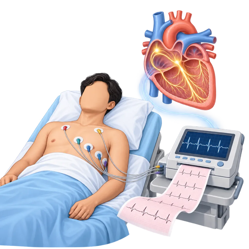
An electrocardiogram, or ECG, is a test that records the heart's electrical activity through electrodes placed on the chest, arms, and legs. The results help your doctor recognize rhythm problems, signs of a heart attack, and other heart abnormalities. The test is painless and takes about 5 to 10 minutes, with the recording itself lasting only about 10 seconds.
Sources01 / Understanding the procedure
About Electrocardiogram
What Is an Electrocardiogram
Every heartbeat is triggered by a small electrical signal that spreads from the upper chambers to the lower chambers of the heart. An electrocardiogram, abbreviated ECG, records this signal through electrodes, which are small conductive stickers attached to the skin. The result is a wavy line graph printed on paper or shown on a screen. The ECG is a basic heart test that is almost always done when someone has their heart checked for the first time.
How It Works in Brief
A standard ECG uses 10 electrodes: six on the chest and four on the arms and legs. From these electrodes, the machine builds 12 leads, which are 12 views of the heart's electrical activity. Each heartbeat appears as a series of waves: the P wave from the upper chambers, the QRS complex as the lower chambers begin to pump, and the T wave as the lower chambers get ready for the next beat. Your doctor assesses the rate, regularity, shape, and spacing of these waves.
What It Can Show
- Rhythm problems: a heartbeat that is too fast, too slow, or irregular, for example atrial fibrillation, as well as electrical conduction problems such as heart block.
- Signs of a heart attack: changes in wave shape that indicate the heart muscle is not getting enough blood, whether it is happening now or happened in the past.
- Thickening or enlargement of the heart: for example due to long-standing hypertension or valve disease.
- Effects of medicines and electrolytes: for example potassium levels that are too high or too low, or medicines that prolong the QT interval.
Limits of This Test
An ECG only records a few seconds. Rhythm problems that come and go may not be captured, so a normal result does not necessarily rule out heart disease. Narrowing of the coronary arteries is also often not visible at rest. For this reason, your doctor may add a longer recording, covered on the Holter Monitoring page, or a test while the heart works harder, covered on the Treadmill Stress Test page.
When an ECG Is Done
An ECG is done when there are symptoms such as chest pain, palpitations, shortness of breath, dizziness, or fainting. For chest pain suspected to be a heart attack, guidelines recommend an ECG within 10 minutes of the patient's arrival. An ECG is also used to monitor heart disease and the effects of medicines, and before surgery in people with heart risk. In people without symptoms and at low risk, a routine ECG is not always needed; your doctor considers it based on age and risk factors, which is covered on the Cardiac Medical Check-Up page.
02 / Understanding the procedure
When Is This Procedure Considered?
A doctor may consider this procedure in the situations below. The decision is always made after a thorough assessment.
- Chest PainTo look for signs of a heart attack or heart muscle that is not getting enough blood flow.
- Palpitations or an Irregular HeartbeatTo identify the type of rhythm problem, for example atrial fibrillation, if it occurs during the recording.
- Fainting, Dizziness, or Shortness of BreathAs an initial test to look for causes that come from the heart.
- Monitoring Heart Disease and MedicinesFor example in heart failure or with medicines that affect the heart's rhythm and electrical conduction.
- Before Surgery or a ProcedureIn people with heart disease or risk factors, based on the doctor's assessment.
- Heart Health CheckupFor people with risk factors such as hypertension, diabetes, or a family history of heart disease.
Situations to Consider First
- Severe Ongoing Chest PainGo to the emergency room, not an outpatient appointment, so the ECG and treatment can be done right away.
- Adhesive Allergy or Sensitive SkinThe type of electrode can be adjusted; tell the staff before the test.
- Wounds or Skin Infection on the ChestThe electrode position can be moved slightly away from the affected skin.
- Difficulty Lying StillMovement, shivering, or coughing interferes with the recording; the staff will help you find a comfortable position.
03 / Before the procedure
How Should You Prepare?
An ECG does not require fasting or any special preparation.
Clothing. Wear a top that is easy to remove or buttons at the front. Necklaces and watches may need to be removed briefly.
Chest skin. Avoid lotion, oil, or powder on your chest on the day of the test so the electrodes stick well. Thick chest hair may need to be shaved slightly.
Medicines. Take your medicines as usual unless your doctor tells you otherwise, and bring a list of the medicines you take.
Previous ECGs. If you have them, bring your previous ECG results so your doctor can compare any changes.
Implanted heart device. Tell the staff if you have a pacemaker or ICD.
Unsure about the preparation instructions? Ask our team before the procedure.
04 / During the procedure
How Is the Procedure Done?
An ECG is done in an examination room, the emergency room, or a hospital ward, without anesthesia and without needles. The whole test takes about 5 to 10 minutes, while the recording itself takes only about 10 seconds.
Preparation in the Examination Room
You will be asked to remove the clothing on your upper body and lie on your back. The staff cleans the skin where the electrodes will be attached.
Attaching the Electrodes
Six electrodes are attached to the chest and four to the wrists and ankles or near them, then connected to the ECG machine with wires. The electrodes only record; no electricity is sent into your body.
Recording
You lie still, breathe normally, and do not talk for a few seconds while the machine records. If the recording is disturbed by movement, it is repeated.
Reading the Results
The machine prints or saves the 12-lead tracing. Your doctor reads the results together with your symptoms and other tests, then explains the next steps.
05 / Benefits and risks
Benefits and Risks Should Be Discussed Together.
Every medical procedure has benefits and risks. Your doctor will explain both for your situation before the procedure.
Benefits You Can Expect
- Quick and PainlessThe recording takes only a few seconds, with no needles, no anesthesia, and no radiation exposure.
- Helps Guide Decisions During Chest PainAn ECG in the first minutes helps your doctor recognize a heart attack that needs treatment right away.
- Identifies the Type of Rhythm ProblemFor example atrial fibrillation, heart block, or extra beats, if they occur during the recording.
- Comparison for Future TestsPrevious recordings help your doctor see changes over time.
The benefits of a procedure are not a promise of a cure. Ask how the results will affect your care plan.
Risks You Should Know
- Mild Skin IrritationFrom the electrode adhesive; usually goes away on its own within a few hours.
- Rhythm Problems Not CapturedProblems that come and go may not appear during the few seconds of recording.
- A Normal Result Does Not Always Mean No DiseaseNarrowing of the coronary arteries is often not visible on an ECG taken at rest.
- Findings That Need ConfirmationSome ECG changes are not specific and need to be confirmed with other tests.
Risks can differ from person to person. A full explanation and consent are given after an individual assessment.
06 / After the procedure
What Is Recovery Like?
There is no recovery period. The electrodes are removed and you can return to your usual activities right away.
Skin. Sticky marks or mild redness from the electrodes usually go away on their own within a few hours.
Results. Your doctor can usually read the ECG results on the same day. A normal result does not always rule out heart disease, so your doctor may suggest further tests based on your symptoms.
Keep your results. Previous recordings are helpful for comparison at future tests, especially if you ever have chest pain.
Warning signs to watch for. Go to the emergency room right away if you experience:
- Chest pain lasting more than a few minutes, especially if it spreads to the arm, neck, or jaw
- Severe or sudden shortness of breath
- Fainting or near fainting
- A very fast heartbeat with dizziness or weakness
Know the Signs That Need Prompt Care.
Follow the team's guidance on warning signs. If severe or sudden symptoms appear after the procedure, go to the nearest emergency department right away or call Heart Attack Center 1500 258.
07 / Team and support
Doctors and Procedure Support
The doctors below are listed for this procedure. Listed by name, not by ranking.

Dr. med. dr. Denio A. Ridjab, Sp.JP(K)
Interventional Cardiology
Coronary Intervention Procedures +2 centers
Clinical focusInterventional Cardiology · Coronary Artery Disease
View Profile
Dr. dr. Jajang Sinardja, Sp.JP(K)
Interventional Cardiology
Coronary Intervention Procedures
Clinical focusAcute Coronary Syndrome · Primary Coronary Intervention
View Profile
dr. Sunu B. Raharjo, Sp.JP(K), Ph.D.
Arrhythmia and Electrophysiology
Arrhythmia and Device Center
Clinical focusClinical and Interventional Electrophysiology · Cardiac Arrhythmia Ablation
View Profile08 / Common questions
Before You Plan the Procedure.
Frequently asked questions about the electrocardiogram (ECG) test.
Is an ECG painful or dangerous?
No. An ECG only records the electrical signals produced by your own heart; no electricity is sent into your body, and there are no needles and no radiation. All you may feel is coldness when the electrodes are attached and slight discomfort when they are removed. An ECG is safe for children, during pregnancy, and for people who have a pacemaker.
How long does an ECG take?
The whole test is usually done in 5 to 10 minutes, including changing clothes and attaching the electrodes. The recording itself takes only about 10 seconds. In the emergency room, an ECG for chest pain is done as soon as possible, within 10 minutes of the patient's arrival according to guidelines, because the results determine the next treatment.
Do I need to fast or stop taking my medicines?
You do not need to fast. Medicines are generally taken as usual, unless your doctor tells you otherwise. Avoid lotion or oil on your chest on the day of the test, wear a top that is easy to remove, and bring a list of your medicines and any previous ECG results so your doctor can compare them.
If my ECG result is normal, does that mean my heart is healthy?
Not necessarily. An ECG only records a few seconds while you are at rest. Rhythm problems that come and go may not be captured, and narrowing of the coronary arteries often only shows up when the heart works harder. If your symptoms continue, your doctor may suggest a Holter, a treadmill test, echocardiography, or other tests based on your symptoms.
What is the difference between an ECG, a Holter, and echocardiography?
An ECG records the heart's electrical activity for a few seconds in the examination room. A Holter records the heart's electrical activity continuously for 24 to 48 hours while you go about your activities, for symptoms that come and go. Echocardiography is different: it is an ultrasound test that shows the shape, chambers, valves, and pumping motion of the heart. The three often complement each other.
Is an ECG from a smartwatch the same as an ECG at the hospital?
No. A smartwatch generally records a single lead for about 30 seconds when you touch it, while a hospital ECG records 12 leads. A smartwatch can give a clue to atrial fibrillation, but it cannot be used to rule out a heart attack. If your smartwatch gives an alert, bring the recording to your consultation; the diagnosis is still confirmed by your doctor with an ECG.
Sources and Medical Review
Translated from the Indonesian edition medically reviewed by dr. Sunu B. Raharjo, Sp.JP(K), Ph.D..
Sources as cited in the Indonesian edition.
- Kligfield P, Gettes LS, Bailey JJ, et al. Recommendations for the standardization and interpretation of the electrocardiogram: part I: the electrocardiogram and its technology. Circulation. 2007;115(10):1306-1324.
- Byrne RA, Rossello X, Coughlan JJ, et al. 2023 ESC Guidelines for the management of acute coronary syndromes. Eur Heart J. 2023;44(38):3720-3826.
- Gulati M, Levy PD, Mukherjee D, et al. 2021 AHA/ACC/ASE/CHEST/SAEM/SCCT/SCMR Guideline for the Evaluation and Diagnosis of Chest Pain: A Report of the American College of Cardiology/American Heart Association Joint Committee on Clinical Practice Guidelines. Circulation. 2021;144(22):e368-e454.
- Van Gelder IC, Rienstra M, Bunting KV, et al. 2024 ESC Guidelines for the management of atrial fibrillation developed in collaboration with the European Association for Cardio-Thoracic Surgery (EACTS). Eur Heart J. 2024;45(36):3314-3414.
- Curry SJ, Krist AH, Owens DK, et al. Screening for Cardiovascular Disease Risk With Electrocardiography: US Preventive Services Task Force Recommendation Statement. JAMA. 2018;319(22):2308-2314.
- Halvorsen S, Mehilli J, Cassese S, et al. 2022 ESC Guidelines on cardiovascular assessment and management of patients undergoing non-cardiac surgery. Eur Heart J. 2022;43(39):3826-3924.