
Quick answer
What Is Ebstein Anomaly?
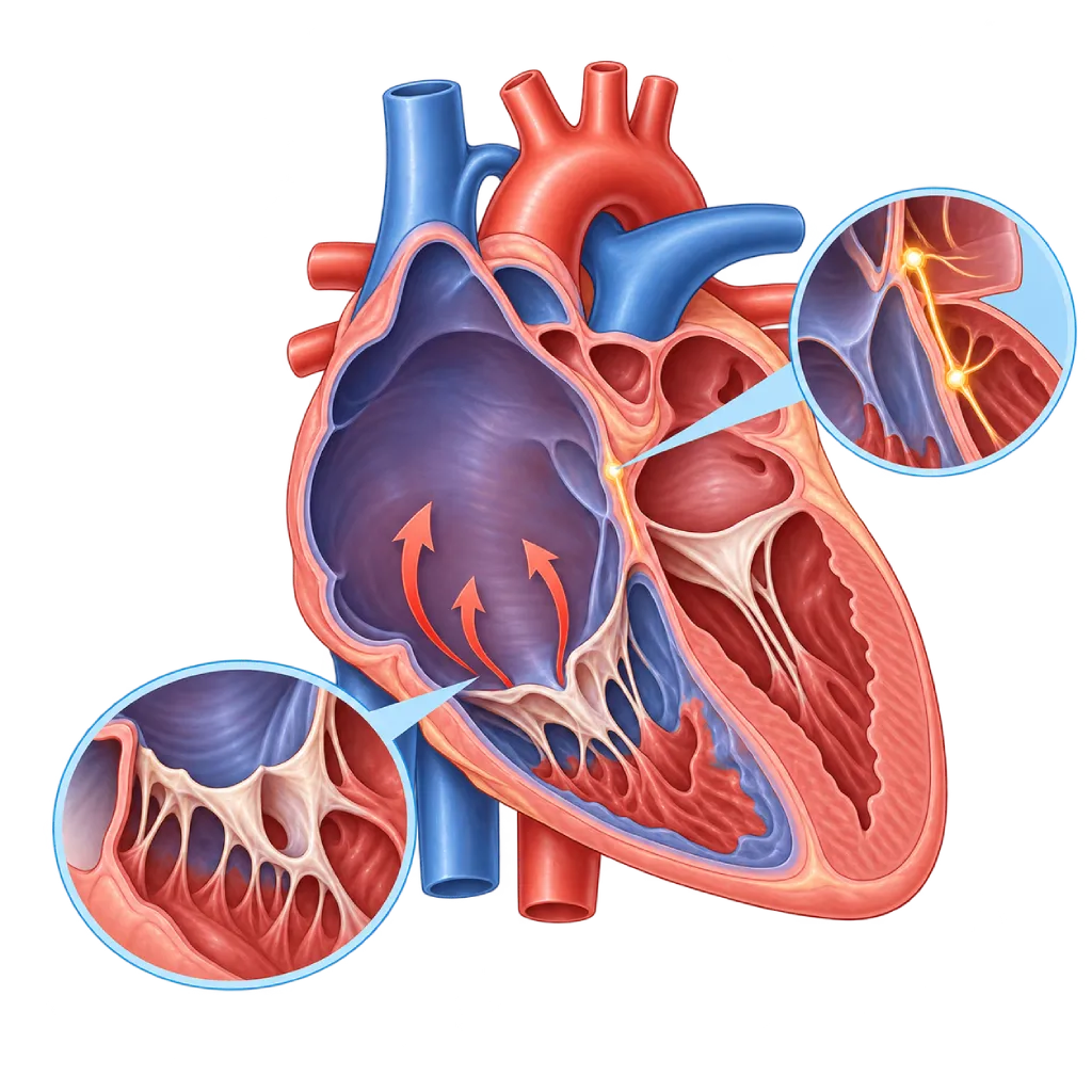
Ebstein anomaly is a congenital defect of the tricuspid valve, the valve between the right atrium and right ventricle, which is displaced downward so the valve leaks and the right side of the heart enlarges. Its severity varies widely.
SourcesSigns That Need Urgent Help
- A very fast heartbeat with dizziness, chest pain, or shortness of breath
- Fainting, especially during or after physical activity
- A bluish color that is getting worse
- A baby who is very short of breath, refuses to feed, or is very limp
- Sudden facial drooping, slurred speech, or weakness on one side of the body
- Rapidly increasing swelling of the legs and abdomen with shortness of breath
If these signs appear, go to the nearest emergency department right away or call Heart Attack Center 1500 258.
01 / Understanding the condition
About Ebstein Anomaly
The tricuspid valve sits between the right atrium and the right ventricle. It keeps blood flowing in one direction into the right ventricle and then on to the lungs. In Ebstein anomaly, the tricuspid valve leaflets form abnormally and attach lower down, inside the right ventricle. As a result, part of the right ventricle becomes part of the atrium, and the pumping chamber of the right ventricle becomes smaller. The valve also does not close tightly, so blood leaks back into the right atrium. Ebstein anomaly is a rare congenital heart defect, accounting for less than 1 percent of all congenital heart disease.
Commonly Associated Defects
Most people with Ebstein anomaly also have a hole in the wall between the atria, either a patent foramen ovale or an atrial septal defect. When pressure in the right atrium is high, oxygen-poor blood crosses to the left side, so the lips and nails look bluish. Some patients are also born with an extra electrical pathway, as in Wolff-Parkinson-White syndrome, which can trigger sudden episodes of a very fast heartbeat. For this reason, a heart rhythm assessment is always part of the evaluation of Ebstein anomaly.
Severity Varies Widely
In severe forms, newborns may look blue and short of breath because blood has trouble flowing to the lungs. The baby's condition often improves once the pressure in the lungs falls over a few days to weeks. In mild forms, a child may grow normally and the condition may only be discovered in the teenage or adult years because of palpitations, tiring easily, or a heart that looks enlarged on an X-ray. Treatment is tailored to age, symptoms, the severity of the leak, the size and function of the right ventricle, oxygen levels, and rhythm disturbances.
Valve Repair
Besides medication and treatment of rhythm disturbances, a valve repair operation called cone reconstruction (cone repair) has been developed over the past two decades. This technique reshapes the patient's own valve leaflets into a cone so they close more tightly, without an artificial valve. If the valve cannot be repaired, it is replaced with an artificial valve. The 2020 ESC and 2018 AHA/ACC guidelines recommend surgery when severe leakage is accompanied by symptoms, or when the right ventricle keeps enlarging or weakening, before the damage becomes permanent.
02 / Understanding the condition
Symptoms to Recognize.
Common Symptoms
- Often no symptoms in mild forms
- Tiring quickly and a reduced ability to be physically active
- Shortness of breath during physical activity
- A very fast heartbeat that starts and stops suddenly
- Bluish lips and nails, especially during physical activity
- Swollen legs or an enlarged abdomen in advanced stages
Symptoms in Newborns
In severe forms, symptoms appear in the first days of life and often improve once the pressure in the baby's lungs falls.
- Bluish skin and lips from birth
- Fast or labored breathing
- Difficulty feeding and tiring quickly
- Looking limp and pale
Symptoms in Children
- Tiring more quickly during play than other children of the same age
- Sudden palpitations that make the child pale or dizzy
- A mild bluish color after running
- A heart murmur heard during a routine checkup
03 / Understanding the condition
What Causes This Condition?
- The tricuspid valve leaflets fail to separate from the wall of the right ventricle while the fetal heart is forming
- In most patients, the exact trigger is unknown
- Changes in certain genes, for example MYH7 or NKX2-5, in some families
- Lithium use in early pregnancy, although the link is now considered weaker than previously thought
- Other congenital heart defects that occur alongside it, such as a hole in the wall between the atria
04 / Understanding the condition
Who Is at Higher Risk?
Genetic
A family history of congenital heart disease and changes in certain genes, such as MYH7, which is also linked to heart muscle disorders, increase the likelihood of Ebstein anomaly.
Medical
Use of certain medications in early pregnancy, particularly lithium for bipolar disorder, has been linked to Ebstein anomaly. Do not stop your medication on your own; discuss it with your doctor before becoming pregnant.
Age
In mild forms, the risk of rhythm disturbances, cyanosis, and a weakening right ventricle increases with age if the condition is not monitored.
Groups at Higher Risk
- Babies with Severe Forms: Babies who are blue from birth and have a severely leaking valve may need intensive care and procedures in the first months of life.
- People with Extra Electrical Pathways: They are at risk of episodes of a very fast heartbeat, so ablation is often recommended before or during surgery.
- Women Planning a Pregnancy: Pregnancy adds to the workload of the right side of the heart and can worsen cyanosis or rhythm disturbances, so a consultation beforehand is needed.
05 / Understanding the condition
How Is It Diagnosed?
In babies, Ebstein anomaly is often suspected because of a bluish color and a heart that looks large on a chest X-ray. Some cases are already visible before birth on fetal echocardiography. In children and adults, clues may include a heart murmur, episodes of palpitations, or a characteristic pattern on the ECG.
Echocardiography confirms the diagnosis by showing the displacement of the tricuspid valve leaflets, the severity of the leak, the size of the right heart chambers, and any hole in the wall between the atria. Cardiac MRI measures the size and function of the right ventricle in detail, which helps determine the timing of surgery. An ECG, Holter monitoring, and, if needed, an electrophysiology study are used to look for an extra electrical pathway. An exercise test assesses physical capacity and any drop in oxygen during activity, which also informs the decision about surgery.
Echocardiography
The main test to see the position of the valve leaflets, the severity of the leak, the size of the right atrium and ventricle, and any hole in the wall between the atria.
Electrocardiogram (ECG)
Looks for an extra electrical pathway, right bundle branch block, and other rhythm disturbances.
Cardiac MRI
Measures the size and function of the right ventricle in detail, helping determine the timing of surgery in teenagers and adults.
Holter Monitoring
Records the heart rhythm for 24 to 48 hours to catch rhythm disturbances that do not show up on a short ECG.
Electrophysiology Study
Confirms the location of the extra electrical pathway and is also part of the ablation procedure.
Treadmill Stress Test
An exercise test to objectively assess physical capacity and any drop in oxygen levels during activity.
Chest X-Ray
Can show a greatly enlarged heart, sometimes the first clue.
06 / Understanding the condition
Treatment Options.
Treatment is decided by a doctor after an assessment. The categories below can help you follow the conversation with your doctor.
Regular Monitoring
Mild forms without symptoms, with a right ventricle that is still working well and no rhythm disturbances, can simply be monitored with regular echocardiography and ECGs.
Medications
Diuretics and rhythm-control medications can relieve swelling and palpitations. In babies with severe forms, certain medications temporarily help blood flow to the lungs. Your doctor determines the type and dose.
Catheter Ablation
The extra electrical pathway or other sources of rhythm disturbances are eliminated through a catheter. Covered on the Cardiac Ablation page.
Tricuspid Valve Repair Surgery
Cone reconstruction (cone repair) reshapes the patient's own valve so it closes more tightly. If this is not possible, the valve is replaced with an artificial valve. The hole in the wall between the atria is usually closed at the same time.
Staged Surgery for Severe Forms
In babies or patients with a very small right ventricle, doctors may choose staged surgery that sends part of the blood directly to the lungs.
Pacemaker or ICD
Considered when there is a disorder of the heart's electrical conduction or a risk of dangerous rhythm disturbances.
07 / Understanding the condition
Complications to Watch For.
Heart Rhythm Disorders
An extra electrical pathway and an enlarged right atrium can trigger supraventricular tachycardia, flutter, or atrial fibrillation. Covered on the Wolff-Parkinson-White Syndrome page.
Right Heart Failure
A severe valve leak and a small right ventricle gradually weaken the pumping of the right side of the heart, causing swelling of the legs and abdomen.
Cyanosis and Low Oxygen Levels
Oxygen-poor blood crossing through the hole in the wall between the atria makes the lips and nails bluish and limits physical activity.
Stroke and Brain Abscess
A hole in the wall between the atria allows clots or germs from the veins to pass to the brain, especially in patients with cyanosis.
Sudden Cardiac Arrest
Very fast rhythm disturbances can, although rarely, cause cardiac arrest. Treating the extra electrical pathway helps lower this risk.
08 / Understanding the condition
What Can You Do?
- If you take lithium and are planning a pregnancy, talk with your doctor without stopping the medication on your own
- Ask about fetal echocardiography if there is a family history of congenital heart disease
- If you have Ebstein anomaly, keep to your follow-up schedule even if you feel well
- Write down episodes of palpitations, including how long they last and what triggers them, to bring to your checkups
- Keep your teeth and gums clean to lower the risk of heart infection
- Ask your doctor which types of exercise are safe for you
09 / Understanding the condition
When Should You See a Doctor?
People with Ebstein anomaly need regular checkups with a doctor experienced in congenital heart disease. Make an appointment sooner if any of the following changes appear. Go to the emergency room right away if you have a very fast heartbeat with dizziness or fainting, or if a baby looks very blue and short of breath.
- Episodes of palpitations that are more frequent or last longer
- Tiring quickly or getting short of breath during activities that used to feel easy
- Increasing bluish color of the lips or nails
- Swollen legs or an enlarged abdomen
- A baby who has difficulty feeding or is not gaining weight well
- You have Ebstein anomaly and are planning a pregnancy
Don't Wait in an Emergency.
If symptoms are severe or come on suddenly, go to the nearest emergency department right away or call Heart Attack Center 1500 258, rather than waiting for an appointment.
10 / Common questions
Questions You May Have.
Frequently asked questions about Ebstein anomaly.
Does Ebstein anomaly always need surgery?
No. Many people with mild forms live for years without surgery, with regular monitoring. The 2020 ESC and 2018 AHA/ACC guidelines recommend surgery when severe valve leakage comes with symptoms, physical capacity declines, cyanosis increases, the right ventricle keeps enlarging or weakening, or a stroke occurs because of blood shunting through the heart. The right timing for surgery is determined from echocardiography, cardiac MRI, and an exercise test.
What is cone reconstruction (cone repair)?
Cone reconstruction is a surgical technique for repairing the tricuspid valve using the patient's own valve tissue. The valve leaflets are freed from the wall of the right ventricle, then reshaped into a cone and stitched into a more normal position. If the repair is successful, the patient does not need an artificial valve. Whether this technique is suitable is assessed from the shape of the valve on echocardiography and cardiac MRI.
Why do people with Ebstein anomaly often have palpitations?
There are two main reasons. First, some patients are born with an extra electrical pathway, as in Wolff-Parkinson-White syndrome, which can trigger a very fast heartbeat. Second, an enlarged right atrium is prone to flutter or atrial fibrillation. The extra pathway can usually be eliminated with catheter ablation. Note the time and duration of each episode, then tell your doctor at your checkup.
Is pregnancy possible with Ebstein anomaly?
Many women with mild to moderate Ebstein anomaly can go through pregnancy well. The risk is higher if there is cyanosis, heart failure, or rhythm disturbances that are not yet under control. ESC guidelines recommend a consultation before pregnancy to assess heart function, treat rhythm disturbances first, and plan monitoring together with an obstetrician. Fetal echocardiography is also recommended.
Can children with Ebstein anomaly play sports?
In mild forms without cyanosis or rhythm disturbances, most children can exercise at light to moderate intensity. Strenuous competitive sports need to be assessed first with an exercise test and rhythm monitoring. If there is an extra electrical pathway, the doctor may recommend ablation before the child becomes active in sports. Discuss suitable types of exercise with your child's doctor.
Sources and Medical Review
Translated from the Indonesian edition medically reviewed by dr. Radityo Prakoso, Sp.JP(K).
Sources as cited in the Indonesian edition.
- Baumgartner H, De Backer J, Babu-Narayan SV, et al. 2020 ESC Guidelines for the management of adult congenital heart disease. Eur Heart J. 2021;42(6):563-645.
- Stout KK, Daniels CJ, Aboulhosn JA, et al. 2018 AHA/ACC Guideline for the Management of Adults With Congenital Heart Disease: A Report of the American College of Cardiology/American Heart Association Task Force on Clinical Practice Guidelines. Circulation. 2019;139(14):e698-e800.
- Holst KA, Connolly HM, Dearani JA. Ebstein's anomaly. Methodist Debakey Cardiovasc J. 2019;15(2):138-44.
- Attenhofer Jost CH, Connolly HM, Dearani JA, Edwards WD, Danielson GK. Ebstein's anomaly. Circulation. 2007;115(2):277-85.
- Holst KA, Dearani JA, Said S, et al. Improving results of surgery for Ebstein anomaly: where are we after 235 cone repairs? Ann Thorac Surg. 2018;105(1):160-8.
- Walsh EP. Ebstein's anomaly of the tricuspid valve: a natural laboratory for re-entrant tachycardias. JACC Clin Electrophysiol. 2018;4(10):1271-88.
Doctors Who Treat This Condition

dr. Salomo Purba, Sp.BTKV, Subsp.JPK(K)
Pediatric and Congenital Heart Surgery
Congenital and Structural Heart Center +2 centers
Clinical focusPediatric Heart Surgery · Congenital Heart Defect Surgery
View Profile
dr. Radityo Prakoso, Sp.JP(K)
Pediatrics & Congenital Heart Disease
Congenital and Structural Heart Center
Clinical focusPediatric Cardiology Consultation · Congenital Heart Disease Care
View Profile
dr. Sunu B. Raharjo, Sp.JP(K), Ph.D.
Arrhythmia and Electrophysiology
Arrhythmia and Device Center
Clinical focusClinical and Interventional Electrophysiology · Cardiac Arrhythmia Ablation
View Profile