
Quick answer
What Is Electrophysiology Study?
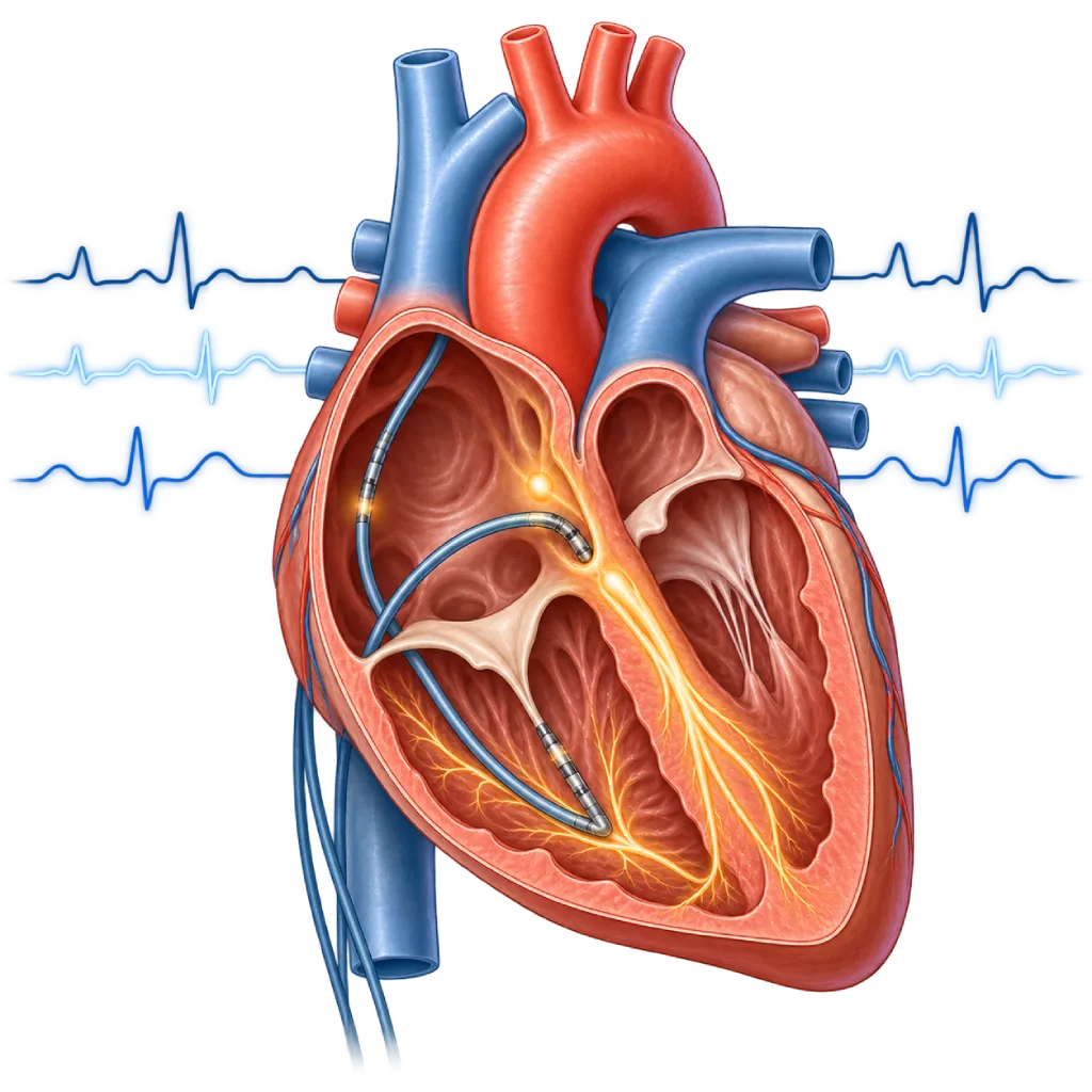
An electrophysiology study tests the heart's electrical system from within. Thin catheters are passed through a vein in the groin to record electrical signals and trigger arrhythmias in a controlled way, so their type and source can be confirmed. The test takes about 1 to 2 hours and is often followed by ablation in the same session.
Sources01 / Understanding the procedure
About Electrophysiology Study
What Is an Electrophysiology Study
An electrophysiology study, often shortened to EPS, is a test that assesses the heart's electrical system from the inside. An ECG and Holter monitoring record the rhythm from the skin surface and only capture heart rhythm disorders (arrhythmias) that happen to occur during the recording. In an EPS, the doctor records signals directly at several points inside the heart and can trigger arrhythmias in a controlled way, so their type and source can be confirmed.
How It Works in Brief
Several thin catheters with electrode tips are passed through a vein in the groin and positioned in the right atrium, near the main conduction pathway, and in the right ventricle. The catheters record how long signals take to travel from the atria to the ventricles. The doctor then delivers small electrical stimuli in specific patterns to see whether there is an extra pathway, a looping circuit, or a spot that triggers a fast heartbeat.
When This Test Is Needed
An EPS is considered when a fast heartbeat that comes and goes has not been clearly recorded, especially if ablation is planned. The test is also used to look for the cause of fainting in people who have had a heart attack or have certain conduction disorders on their ECG, to tell whether a fast rhythm comes from the atria or the ventricles, and to assess the risk of an extra pathway, which is covered on the Wolff-Parkinson-White Syndrome page.
Often Followed by Ablation
If the source of the arrhythmia is found and can be treated, the doctor usually performs ablation right away in the same session, without a new puncture. For this reason, consent for a possible ablation is usually discussed before the test. The treatment procedure is covered on the Cardiac Ablation page.
What the Results Mean
The EPS results help the doctor choose the next step. If the arrhythmia can be triggered and its source is clear, ablation becomes an option. If conduction from the atria to the ventricles is very slow, the doctor may recommend a pacemaker, which is covered on the Pacemaker Implantation page. If no abnormality is found, a longer rhythm recording may be recommended, which is covered on the Long-Term Heart Rhythm Monitoring page.
The decision to implant an ICD is based mainly on the strength of the heart's pump, the history of arrhythmias, and the underlying disease, not only on the EPS results. Comprehensive arrhythmia treatment is covered on the Arrhythmia Management page.
02 / Understanding the procedure
When Is This Procedure Considered?
A doctor may consider this procedure in the situations below. The decision is always made after a thorough assessment.
- Fast Palpitations That Come and GoSuspected supraventricular tachycardia that has not been clearly recorded, especially if ablation is planned.
- Wolff-Parkinson-White SyndromeTo assess the risk of the extra pathway in certain patients, including those who have never had palpitations.
- Fainting After a Heart AttackFainting without a clear cause in a heart with scar tissue, for example from a previous heart attack.
- Fainting with a Conduction DisorderFor example, bundle branch block on the ECG, to assess whether a pacemaker is needed.
- Fast Rhythm of Unclear OriginTo tell whether a fast rhythm comes from the atria or the ventricles when the ECG cannot confirm it.
Situations to Consider First
- Ongoing Heart Attack or Chest PainReduced blood flow to the heart muscle is treated first, before the test.
- Worsening Heart FailureThe test is postponed until shortness of breath and swelling are controlled with medicines.
- Active Infection or FeverThe test is postponed until the infection is treated so that germs do not spread to the heart.
- Electrolyte Imbalance or Medicine EffectsFor example, low potassium or an excess of certain medicines, which needs to be corrected first.
- PregnancyUsually postponed because of X-ray exposure, unless the benefit is clearly greater.
03 / Before the procedure
How Should You Prepare?
An EPS is usually scheduled. The team explains the preparation and asks for your consent, including for a possible ablation in the same session.
Fasting. Usually no food or drink for about 6 hours before the test.
Heart rhythm medicines. Medicines such as beta blockers or antiarrhythmic drugs may need to be stopped a few days beforehand so the arrhythmia can be triggered easily. Follow the schedule from your doctor and do not stop any medicine without instructions.
Blood thinners and diabetes medicines. Their doses may be adjusted on the day of the test; ask the team for instructions.
Symptom recordings. Bring ECG or Holter results, or smartwatch recordings taken during palpitations. These recordings help the doctor match the triggered arrhythmia with your symptoms.
Information for the team. Tell the team about any allergies, whether you might be pregnant, and your medical history. Bring a family member to accompany you and take you home.
Unsure about the preparation instructions? Ask our team before the procedure.
04 / During the procedure
How Is the Procedure Done?
An EPS is done in the electrophysiology laboratory and takes about 1 to 2 hours, longer if it is followed by ablation. The groin is numbed with local anesthesia. A mild sedative may be given, but the dose is limited so it does not suppress the arrhythmia that needs to be triggered, so you stay awake and can talk with the team.
Placing the Catheters
The doctor places a small tube in the groin vein, then advances two to four catheters to the heart under X-ray guidance. You do not feel the catheters moving inside the blood vessels.
Recording Baseline Signals
The doctor measures how long signals take to travel from the atria to the ventricles and assesses the function of the sinus node, the heart's natural pacemaker.
Triggering the Arrhythmia
The heart is paced with small electrical stimuli at various speeds. You may feel palpitations similar to your usual symptoms. A stimulating medicine may be given through an IV line if the arrhythmia is hard to trigger. If a fast rhythm does not stop on its own, the team stops it with pacing or an electrical shock after giving you medicine to put you to sleep briefly.
Deciding the Next Step
If the source of the arrhythmia is found and you have agreed beforehand, ablation can be done right away. When the test is finished, the catheters are removed and the groin is pressed to prevent bleeding.
05 / Benefits and risks
Benefits and Risks Should Be Discussed Together.
Every medical procedure has benefits and risks. Your doctor will explain both for your situation before the procedure.
Benefits You Can Expect
- The Type and Source of the Arrhythmia Become ClearSignals are recorded directly from inside the heart, not only from the skin surface.
- Can Be Followed Directly by AblationIf the source is found, treatment can be done in the same session.
- Helps Decide on a PacemakerIn fainting with a conduction disorder, the results help determine whether a device is needed.
- No IncisionThe catheters enter through a small puncture in the groin, and recovery is short.
The benefits of a procedure are not a promise of a cure. Ask how the results will affect your care plan.
Risks You Should Know
- Bruising or Bleeding in the GroinUsually mild and improves on its own within a few days.
- Blood Vessel InjuryRare; for example, a pulsating lump or a connection between an artery and a vein that needs treatment.
- Arrhythmia That Needs to Be StoppedA triggered fast rhythm sometimes needs pacing or an electrical shock from the team.
- Fluid or Blood Around the HeartVery rare; if it occurs, the fluid is promptly drained with a needle.
- Blood Clot in a Leg VeinRare; prevented by starting to move gradually once the bed rest period is over.
Risks can differ from person to person. A full explanation and consent are given after an individual assessment.
06 / After the procedure
What Is Recovery Like?
After the test, you are monitored in the recovery room. Many patients stay one night, especially if ablation was also done; some can go home the same day.
Lying down after the test. The leg used for the puncture is kept straight for 2 to 4 hours to prevent bleeding in the groin.
Activity. Light activities can be resumed the next day. Avoid heavy lifting for a few days.
Results. The doctor usually explains the findings on the same day, including whether ablation was done or other treatment is needed, such as medicines, a pacemaker, or a longer rhythm recording.
Medicines. Ask when you can restart any heart rhythm medicines that were stopped.
Warning signs to watch for. Go to the emergency room right away if you experience:
- Bleeding or swelling in the groin that keeps getting bigger
- A cold, pale, or painful leg
- Chest pain, shortness of breath, or fainting
- A fast heartbeat that does not stop
Know the Signs That Need Prompt Care.
Follow the team's guidance on warning signs. If severe or sudden symptoms appear after the procedure, go to the nearest emergency department right away or call Heart Attack Center 1500 258.
07 / Team and support
Doctors and Procedure Support
The doctors below are listed for this procedure. Listed by name, not by ranking.

Dr. dr. Dicky A. Hanafy, Sp.JP(K)
Arrhythmia and Electrophysiology
Arrhythmia and Device Center +1 center
Clinical focusElectrophysiology and Arrhythmia Ablation · Pacemaker and ICD Implantation
View Profile
Dr. dr. Faris Basalamah, Sp.JP(K)
Interventional Cardiology
Coronary Intervention Procedures +1 center
Clinical focusComplex Coronary Intervention · Cardiac Electrophysiology
View Profile
dr. Sunu B. Raharjo, Sp.JP(K), Ph.D.
Arrhythmia and Electrophysiology
Arrhythmia and Device Center
Clinical focusClinical and Interventional Electrophysiology · Cardiac Arrhythmia Ablation
View ProfileHD Grid 3D Mapping System
A mapping catheter made by Abbott with electrodes arranged in a grid, recording the heart's electrical signals from many points…
EnSite X EP System
A 3D mapping system made by Abbott that maps the shape and electrical activity of the heart chambers during electrophysiology…
Electrophysiology (EP) Laboratory
A dedicated procedure room for electrophysiology studies, catheter ablation, and implantation of heart rhythm devices such as…
The availability of technology and test support is confirmed with the team. Technology is chosen according to clinical need.
08 / Common questions
Before You Plan the Procedure.
Frequently asked questions about electrophysiology studies.
Is an electrophysiology study painful?
The puncture site in the groin is numbed with local anesthesia, so you usually feel only the initial needle prick and some pressure. You do not feel the catheters inside the blood vessels and heart. When the heart is paced, you may feel palpitations, dizziness, or chest discomfort similar to your usual symptoms. Tell the team if you feel uncomfortable; more sedative can be given as needed.
Why are my heart rhythm medicines stopped before the test?
Medicines such as beta blockers or antiarrhythmic drugs can suppress the arrhythmia, making it hard to trigger during the test. If the arrhythmia does not appear, its source cannot be found and ablation cannot be done. For this reason, your doctor may ask you to stop the medicines a few days beforehand. Do not stop them on your own, because some medicines need to be continued or stopped gradually.
What is the difference between an electrophysiology study and cardiac ablation?
An electrophysiology study is a test to find the type and source of an arrhythmia. Ablation is a procedure to deactivate that electrical source or pathway. Both use the same catheters and are often done one after the other in a single session. In some patients, an EPS is done on its own, for example to look for the cause of fainting. Ablation is covered on the Cardiac Ablation page.
What if the arrhythmia cannot be triggered?
This result does not always mean you are free of arrhythmias. Some arrhythmias only appear under certain conditions and are not triggered during the test. The doctor assesses the results together with your symptoms and may recommend a longer rhythm recording, including with a recorder implanted under the skin. This is covered on the Long-Term Heart Rhythm Monitoring and Implantable Loop Recorder (ILR) pages.
Am I awake during the test?
Usually, yes. You are given local anesthesia and a mild sedative, so you can still talk with the team and tell them if your usual symptoms appear. The sedative is limited because it can suppress the arrhythmia. If a triggered arrhythmia needs to be stopped with an electrical shock, you are first given medicine to put you to sleep briefly.
When can I return to my usual activities?
If you only had the test, light activities can usually be resumed the next day and work within 1 to 2 days. Avoid heavy lifting for a few days so the puncture site in the groin can heal. If ablation was also done, recovery may take a little longer. Ask your doctor when you can drive.
Sources and Medical Review
Translated from the Indonesian edition medically reviewed by dr. Sunu B. Raharjo, Sp.JP(K), Ph.D..
Sources as cited in the Indonesian edition.
- Brugada J, Katritsis DG, Arbelo E, et al. 2019 ESC Guidelines for the management of patients with supraventricular tachycardia. Eur Heart J. 2020;41(5):655-720.
- Page RL, Joglar JA, Caldwell MA, et al. 2015 ACC/AHA/HRS Guideline for the Management of Adult Patients With Supraventricular Tachycardia. Circulation. 2016;133(14):e506-e574.
- Brignole M, Moya A, de Lange FJ, et al. 2018 ESC Guidelines for the diagnosis and management of syncope. Eur Heart J. 2018;39(21):1883-1948.
- Glikson M, Nielsen JC, Kronborg MB, et al. 2021 ESC Guidelines on cardiac pacing and cardiac resynchronization therapy. Eur Heart J. 2021;42(35):3427-3520.
- Kusumoto FM, Schoenfeld MH, Barrett C, et al. 2018 ACC/AHA/HRS Guideline on the Evaluation and Management of Patients With Bradycardia and Cardiac Conduction Delay: A Report of the American College of Cardiology/American Heart Association Task Force on Clinical Practice Guidelines and the Heart Rhythm Society. Circulation. 2019;140(8):e382-e482.
- Zeppenfeld K, Tfelt-Hansen J, de Riva M, et al. 2022 ESC Guidelines for the management of patients with ventricular arrhythmias and the prevention of sudden cardiac death. Eur Heart J. 2022;43(40):3997-4126.