
Quick answer
What Is Ventricular Tachycardia?
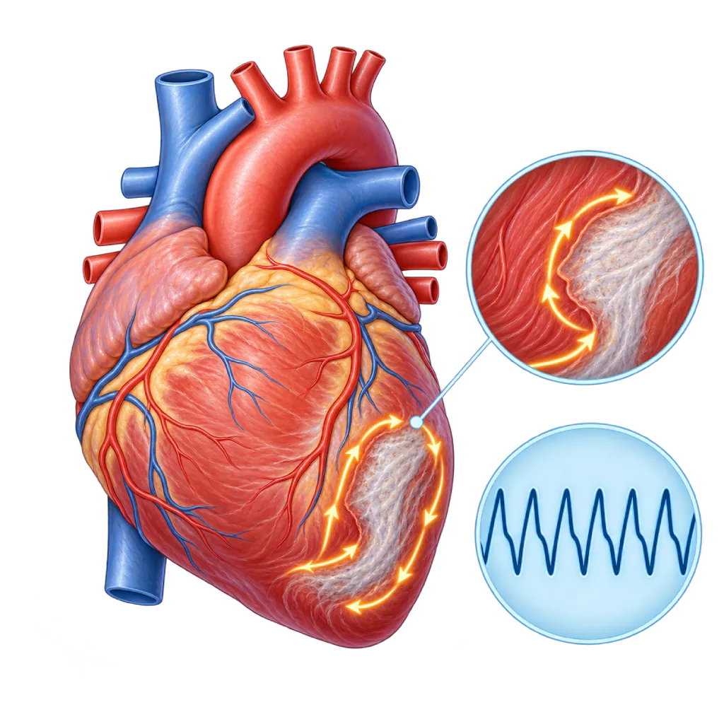
Ventricular tachycardia is a fast heartbeat that starts in the lower chambers of the heart. A long-lasting episode can disrupt the heart's pumping and turn into cardiac arrest, so it needs prompt treatment and prevention of recurrence.
SourcesSigns That Need Urgent Help
- Fainting or loss of consciousness
- Not breathing normally or not responding when called
- Severe chest pain
- Severe shortness of breath
- A very fast heartbeat with paleness, cold sweat, or confusion
- The ICD delivers more than one shock within a short time
If these signs appear, go to the nearest emergency department right away or call Heart Attack Center 1500 258.
01 / Understanding the condition
About Ventricular Tachycardia
Ventricular tachycardia is a rhythm disorder in which the ventricles (the lower chambers of the heart) beat fast on their own signals, usually more than 100 times per minute and often much faster. Normally, the ventricles wait for a signal from the upper chambers. In ventricular tachycardia, signals arise or circle inside the ventricles, so the heart's contractions become less coordinated and less blood is pumped. On an ECG, the waves can look uniform (monomorphic) or keep changing (polymorphic), and this difference helps your doctor look for the cause.
Brief or Sustained
Doctors distinguish nonsustained ventricular tachycardia, which stops on its own in less than 30 seconds, from sustained ventricular tachycardia, which lasts longer or causes blood pressure to drop. The brief form often causes no symptoms and is found on a Holter recording, but its cause still needs to be looked for. The sustained form is an emergency because it can turn into ventricular fibrillation, a chaotic rhythm that makes the heart stop pumping. That condition is covered on the Ventricular Fibrillation page.
Causes to Look For
A common cause is scar tissue in the heart muscle after a heart attack. This scar tissue forms pathways where signals can circle. Cardiomyopathy, inflammation of the heart muscle, and inherited abnormalities of the electrical system, such as long QT syndrome or Brugada syndrome, can also be causes. Low potassium or magnesium levels and certain medicines can trigger episodes. In some people with a healthy heart, ventricular tachycardia comes from a single spot in the outflow tract of a ventricle; this type is usually not life-threatening and can be treated with medicines or ablation.
Long-Term Treatment
After an episode is controlled, your doctor looks for the cause and assesses the heart's pumping strength. Treatment may include medicines, catheter ablation to deactivate the source of the signals, and an implantable cardioverter defibrillator (ICD), which can stop dangerous episodes automatically. The underlying disease, such as coronary artery disease or heart failure, is also treated. If someone in your family died suddenly at a young age, tell your doctor, because this may point to an inherited abnormality.
02 / Understanding the condition
Symptoms to Recognize.
Common Symptoms
- A very fast, strong, pounding heartbeat
- Dizziness or light-headedness
- Shortness of breath
- Chest pain or pressure
- Sudden weakness and sweating
- Nearly fainting
- No symptoms in brief episodes found on a rhythm recording
03 / Understanding the condition
What Causes This Condition?
- Scar tissue in the heart muscle after a heart attack
- Reduced blood flow to the heart muscle due to coronary artery disease
- Cardiomyopathy, including dilated, hypertrophic, and arrhythmogenic cardiomyopathy
- Inflammation of the heart muscle (myocarditis) or cardiac sarcoidosis
- Inherited abnormalities of the electrical system, such as long QT syndrome or Brugada syndrome
- Low blood potassium or magnesium levels
- Medicines or substances that affect the heart's electrical activity, including cocaine and stimulants
- A trigger spot in the outflow tract of a ventricle in a healthy heart
04 / Understanding the condition
Who Is at Higher Risk?
Medical
A history of heart attack, heart failure with a weak pump, cardiomyopathy, myocarditis, and severe valve disease increase the risk of ventricular tachycardia.
Genetic
Inherited cardiomyopathies and ion channel disorders such as long QT syndrome or Brugada syndrome can be passed down in families; a family history of sudden death at a young age should be shared with your doctor.
Lifestyle
Cocaine or stimulant misuse, heavy drinking over a long time, and smoking, which worsens coronary artery disease, increase the risk.
Age
Ventricular tachycardia caused by scar tissue is more common in middle-aged and older adults, while inherited causes often appear in children, teenagers, or young adults.
Other
Electrolyte disturbances, for example from severe diarrhea or diuretic medicines, and medicines that prolong the QT interval can trigger episodes.
Groups at Higher Risk
- People Who Have Had a Heart Attack: Scar tissue after a heart attack becomes a pathway for circling signals, especially when the heart's pumping strength is reduced.
- People with Heart Failure or Cardiomyopathy: Heart muscle that is weak, thickened, or replaced by connective tissue more easily produces dangerous rhythms from the ventricles.
- Families with a History of Sudden Death: Sudden death at a young age in the family may indicate an inherited cardiomyopathy or ion channel disorder.
- People with Ion Channel Disorders: Long QT syndrome, Brugada syndrome, and similar disorders make the heart's electrical system more prone to ventricular tachycardia.
05 / Understanding the condition
How Is It Diagnosed?
During an episode, an ECG shows a fast rhythm with wide ventricular waves, because the signal spreads through the ventricles without using the normal pathway. This recording is important for distinguishing ventricular tachycardia from other types of tachycardia. If episodes are infrequent, a Holter monitor, a long-term recorder, or data from an ICD helps capture them. Blood tests check electrolytes and signs of heart muscle damage.
Once your condition is stable, echocardiography assesses the heart's pumping strength, which helps decide whether an ICD is needed. Cardiac MRI can show scar tissue or inflammation in the heart muscle. Coronary angiography or coronary CT looks for narrowing of the heart's blood vessels. In certain situations, your doctor recommends a treadmill test, genetic testing, or an electrophysiology study to map the source of the signals before ablation.
Electrocardiogram (ECG)
Shows a fast rhythm with wide ventricular waves and helps distinguish ventricular tachycardia from other types of tachycardia.
Holter Monitoring
Captures brief episodes that come and go, and counts how often rhythms from the ventricles appear.
Echocardiography
Assesses the heart's pumping strength (ejection fraction), an important basis for deciding whether an ICD is needed.
Cardiac MRI
Shows scar tissue, inflammation, or cardiomyopathy that serves as the source of circling signals.
Coronary Angiography
Looks for narrowed coronary arteries that can trigger ventricular tachycardia and need to be opened.
Blood Electrolyte Test
Checks potassium and magnesium, which can trigger episodes when they are too low.
Electrophysiology Study
Triggers and maps the source of the tachycardia in a controlled way, especially before or during ablation.
06 / Understanding the condition
Treatment Options.
Treatment is decided by a doctor after an assessment. The categories below can help you follow the conversation with your doctor.
Emergency Treatment
During an episode, the doctor delivers a controlled electric shock (cardioversion or defibrillation) or gives antiarrhythmic medicine through an IV, depending on how stable your blood pressure is.
Treating Causes and Triggers
Correcting potassium and magnesium levels, stopping trigger medicines, and opening blocked coronary arteries when they are the cause.
Antiarrhythmic Medicines and Beta-Blockers
Beta-blockers and antiarrhythmic medicines help prevent repeat episodes. Your doctor decides the type and dose, with monitoring for side effects.
Implantable Cardioverter Defibrillator (ICD)
A device under the skin of the chest that monitors the rhythm continuously and stops ventricular tachycardia with rapid pacing or an electric shock. Recommended after a sustained episode or when the heart's pump is weak.
Catheter Ablation
Deactivates the source or pathway of the signals in the ventricle through a catheter. In repeated ventricular tachycardia caused by scar tissue, ablation can reduce episodes and ICD shocks.
Heart Failure Treatment
Guideline-based heart failure medicines and, in certain patients, resynchronization therapy help strengthen the pump and reduce episodes.
07 / Understanding the condition
Complications to Watch For.
Ventricular Fibrillation and Cardiac Arrest
Sustained ventricular tachycardia can turn into a chaotic rhythm that makes the heart stop pumping. Chest compressions and an AED are needed right away.
Fainting and Injury
A drop in blood pressure during an episode can cause sudden fainting, including while driving or being active.
Heart Failure
Repeated episodes and the same underlying disease can weaken the heart's pump, causing shortness of breath and swelling.
Electrical Storm
Three or more episodes within 24 hours, often with repeated ICD shocks. This situation requires hospital care.
08 / Understanding the condition
What Can You Do?
- Control your blood pressure, cholesterol, and blood sugar, and quit smoking
- Take your heart medicines on schedule and do not stop them on your own
- Ask your doctor before using any new medicine, including over-the-counter medicines and supplements
- Avoid cocaine, stimulants, and heavy drinking
- Have your ICD checked on schedule and learn what to do if the device delivers a shock
- Encourage your family to learn chest compressions and how to use an AED
- Share any family history of sudden death so other family members can be checked
09 / Understanding the condition
When Should You See a Doctor?
Ventricular tachycardia always needs to be evaluated by a cardiologist, including the brief form that causes no symptoms. Make an appointment soon if your Holter or ECG results show a rhythm from the ventricles, or if you have heart disease and start having frequent palpitations. For danger signs, do not wait.
- A fast heartbeat with dizziness or nearly fainting
- Holter or ECG results showing ventricular tachycardia, even if brief
- A past heart attack or heart failure, and palpitations that have become frequent
- A family member who died suddenly at a young age
- Fainting, chest pain, severe shortness of breath, or repeated ICD shocks: go to the emergency room right away
Don't Wait in an Emergency.
If symptoms are severe or come on suddenly, go to the nearest emergency department right away or call Heart Attack Center 1500 258, rather than waiting for an appointment.
10 / Common questions
Questions You May Have.
Frequently asked questions about ventricular tachycardia.
What is the difference between ventricular tachycardia and ventricular fibrillation?
In ventricular tachycardia, the ventricles beat fast but still regularly, so some blood is still pumped and a person may stay conscious. In ventricular fibrillation, the signals in the ventricles are chaotic, so the heart stops pumping and the person loses consciousness immediately. Sustained ventricular tachycardia can turn into ventricular fibrillation, so both are treated as emergencies.
Is brief ventricular tachycardia on a Holter result dangerous?
Nonsustained ventricular tachycardia that stops on its own within a few seconds can be found in people with healthy hearts as well as in those with heart disease. What it means depends on the condition of your heart. That is why your doctor will check the structure and pumping strength of your heart, for example with echocardiography or MRI. If your heart is healthy, the risk is usually low; if there is heart disease, treatment is more intensive.
Does everyone with ventricular tachycardia need an ICD?
Not everyone. An ICD is recommended for people who have had sustained ventricular tachycardia or cardiac arrest without a correctable cause, and for some people with a very weak heart pump. In ventricular tachycardia from a trigger spot in a healthy heart, medicines or ablation are usually enough. Your doctor will explain the considerations based on your test results.
What should I do if my ICD delivers a shock?
If you feel well after a single shock, stay calm and contact your doctor or ICD monitoring team that same day. Go to the emergency room right away if you receive more than one shock within a short time, or if a shock comes with chest pain, shortness of breath, or fainting. Your family should know how to call emergency services and start chest compressions if you are unconscious.
Can ablation get rid of ventricular tachycardia?
In ventricular tachycardia from a single spot in a healthy heart, ablation can often eliminate the source. With scar tissue after a heart attack, ablation usually aims to reduce episodes and ICD shocks, not to replace the ICD. Clinical trial results show that ablation was more beneficial than adding medicines in certain patients. Your doctor will explain the benefits and risks for you.
Can I drive or exercise?
After an episode of ventricular tachycardia or ICD placement, you usually need to stop driving for a while, because fainting while driving puts you and others in danger. Your doctor decides how long based on your condition. Light to moderate exercise is often still recommended once your condition is stable, but the type and intensity should be agreed on with your doctor, especially in inherited cardiomyopathy.
Sources and Medical Review
Translated from the Indonesian edition medically reviewed by dr. Sunu B. Raharjo, Sp.JP(K), Ph.D..
Sources as cited in the Indonesian edition.
- Zeppenfeld K, Tfelt-Hansen J, de Riva M, et al. 2022 ESC Guidelines for the management of patients with ventricular arrhythmias and the prevention of sudden cardiac death. Eur Heart J. 2022;43(40):3997-4126.
- Al-Khatib SM, Stevenson WG, Ackerman MJ, et al. 2017 AHA/ACC/HRS Guideline for Management of Patients With Ventricular Arrhythmias and the Prevention of Sudden Cardiac Death. Circulation. 2018;138(13):e272-e391.
- Cronin EM, Bogun FM, Maury P, et al. 2019 HRS/EHRA/APHRS/LAHRS expert consensus statement on catheter ablation of ventricular arrhythmias. Heart Rhythm. 2020;17(1):e2-e154.
- Sapp JL, Wells GA, Parkash R, et al. Ventricular Tachycardia Ablation versus Escalation of Antiarrhythmic Drugs. N Engl J Med. 2016;375(2):111-121.
- Arbelo E, Protonotarios A, Gimeno JR, et al. 2023 ESC Guidelines for the management of cardiomyopathies. Eur Heart J. 2023;44(37):3503-3626.
Doctors Who Treat This Condition

Dr. dr. Dicky A. Hanafy, Sp.JP(K)
Arrhythmia and Electrophysiology
Arrhythmia and Device Center +1 center
Clinical focusElectrophysiology and Arrhythmia Ablation · Pacemaker and ICD Implantation
View Profile
dr. Sunu B. Raharjo, Sp.JP(K), Ph.D.
Arrhythmia and Electrophysiology
Arrhythmia and Device Center
Clinical focusClinical and Interventional Electrophysiology · Cardiac Arrhythmia Ablation
View Profile
Dr. dr. Faris Basalamah, Sp.JP(K)
Interventional Cardiology
Coronary Intervention Procedures +1 center
Clinical focusComplex Coronary Intervention · Cardiac Electrophysiology
View Profile