
Quick answer
What Is Dilated Cardiomyopathy?
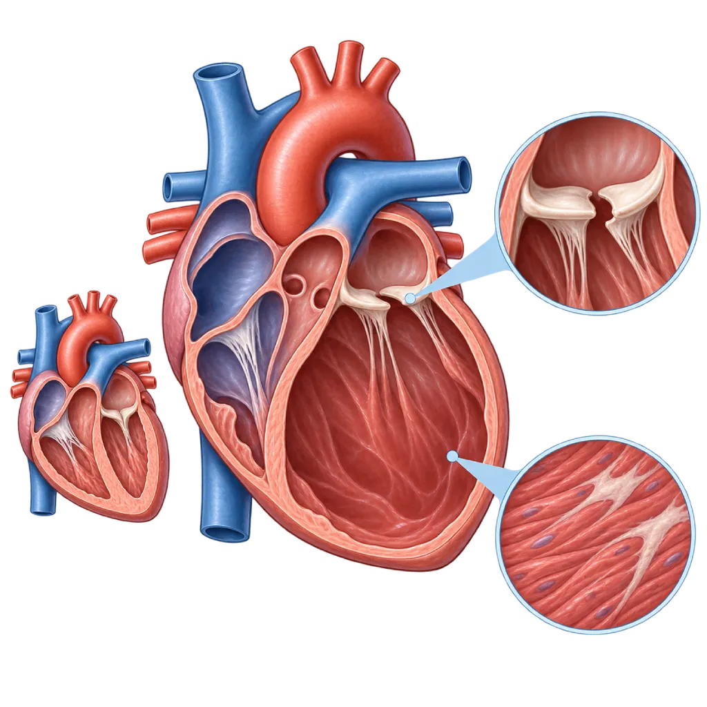
Dilated cardiomyopathy is a heart muscle disease that makes the left ventricle enlarge and its pumping weaken, in a way not sufficiently explained by coronary artery disease or excess workload, and it can lead to heart failure and rhythm disorders.
SourcesSigns That Need Urgent Help
- Severe shortness of breath at rest or needing to sleep sitting up
- Coughing up frothy or pink sputum
- Fainting or nearly fainting
- A very fast heartbeat with weakness or a cold sweat
- Chest pain that does not go away after a few minutes
- Cold, pale skin, confusion, or passing very little urine
If these signs appear, go to the nearest emergency department right away or call Heart Attack Center 1500 258.
01 / Understanding the condition
About Dilated Cardiomyopathy
Dilated cardiomyopathy (DCM) is a condition in which the left ventricle, the heart's main pumping chamber, becomes enlarged and its contraction weakens. According to the 2023 European Society of Cardiology (ESC) guidelines, this diagnosis is used when the enlargement and weakening are not sufficiently explained by coronary artery disease or by excess workload such as hypertension and valve disease. The right ventricle may also become enlarged. As a result, less blood is pumped to the rest of the body and fluid builds up easily in the lungs and legs. This condition can affect both children and adults, and is found more often in men.
A Range of Causes
In quite a few patients, dilated cardiomyopathy is linked to gene changes, for example in the titin (TTN), lamin (LMNA), or filamin C (FLNC) genes, and can be found in more than one family member. Acquired causes include inflammation of the heart muscle (myocarditis), excessive alcohol, chemotherapy drugs such as anthracyclines, rhythm disorders that stay too fast for a long time, pregnancy, thyroid disorders, and deficiencies of certain nutrients. In some people, genes and external triggers act together, while in others no cause is found.
How It Progresses
In the early stages, many people with the condition have no symptoms yet. Symptoms often appear gradually: shortness of breath when climbing stairs, tiring easily, then swelling in the feet. Rhythm disorders can appear first, especially with certain gene changes such as LMNA, so assessing rhythm risk is part of the care. With appropriate heart failure medicines, quite a few patients see an improvement in chamber size and pumping strength within a few months. Repeat echocardiography helps your doctor assess this improvement. Even so, the medicines need to be continued because stopping them can cause the condition to worsen again.
How It Relates to Other Types
Dilated cardiomyopathy is one type within the cardiomyopathy family, which is covered on the Cardiomyopathy page. The term nonischemic cardiomyopathy, often used by doctors, includes dilated cardiomyopathy and other forms of pump weakening not caused by coronary artery disease, and is covered on the Nonischemic Cardiomyopathy page. When the cause is alcohol or medicines, it is covered on the Toxic Cardiomyopathy page, while cardiomyopathy that appears around the time of giving birth is covered on the Peripartum Cardiomyopathy page.
02 / Understanding the condition
Symptoms to Recognize.
Common Symptoms
- Shortness of breath during activity, and later also when lying flat
- Waking at night because of shortness of breath or coughing
- Tiring easily and a reduced ability to be active
- Swelling in the ankles, legs, or abdomen
- Rapid weight gain from fluid buildup
- A pounding heartbeat or a beat that feels irregular
- Dizziness or feeling close to fainting
Symptoms in Children
Dilated cardiomyopathy is a type of cardiomyopathy commonly found in children. Its symptoms often resemble a respiratory infection or a digestive problem.
- Rapid breathing or sweating while feeding or eating
- Difficulty gaining weight as expected for age
- Tiring easily, weakness, or fussiness
- Coughing or wheezing that does not improve
- Nausea, vomiting, or abdominal pain with no clear cause
03 / Understanding the condition
What Causes This Condition?
- Changes in heart muscle genes, for example in the TTN, LMNA, or FLNC genes, which can be inherited
- Myocarditis, or inflammation of the heart muscle, for example after a viral infection
- Excessive alcohol consumption over a long period
- Certain chemotherapy drugs, such as anthracyclines, or radiation to the chest area
- Rhythm disorders that stay too fast for a long period
- Changes around pregnancy and after giving birth (peripartum cardiomyopathy)
- Hormone disorders such as thyroid disease, or deficiencies of certain nutrients
- Autoimmune diseases or inherited muscle diseases that also affect the heart muscle
04 / Understanding the condition
Who Is at Higher Risk?
Genetic
A family history of dilated cardiomyopathy, heart failure at a young age, a pacemaker or ICD implanted at a young age, or sudden cardiac arrest.
Lifestyle
Drinking excessive alcohol for years and using substances such as cocaine or methamphetamine can damage the heart muscle.
Medical
A history of myocarditis, chemotherapy or chest radiation, thyroid disorders, autoimmune disease, and uncontrolled fast rhythm disorders.
Other
Pregnancy and the postpartum period, as well as inherited muscle diseases such as Duchenne or Becker muscular dystrophy, which can also affect the heart.
Groups at Higher Risk
- Family Members of People with Dilated Cardiomyopathy: Dilated cardiomyopathy can be found in more than one family member, so close relatives are advised to be checked even without symptoms.
- Heavy Drinkers: Excessive alcohol consumption over many years can weaken the heart muscle. Pumping function often improves when alcohol is stopped.
- Cancer Survivors: Anthracyclines, trastuzumab, and chest radiation can reduce pumping function, so heart monitoring is recommended during and after treatment.
- Pregnant and Postpartum Women: Peripartum cardiomyopathy has a similar picture and can appear up to several months after delivery.
- People with Muscular Dystrophy: Duchenne and Becker muscular dystrophy are often accompanied by dilated cardiomyopathy, so the heart needs to be checked regularly.
05 / Understanding the condition
How Is It Diagnosed?
Your doctor asks about your symptoms, alcohol use, medicines you have taken including chemotherapy, any history of infection or pregnancy, and your family history. Echocardiography is usually the first test, to see enlargement of the left ventricle and measure the ejection fraction. The next step is to confirm that the weakened pumping is not caused by coronary artery disease, for example with coronary CT or coronary angiography. Cardiac MRI with contrast helps look for scar tissue, inflammation, or damage patterns typical of certain causes. Blood tests are used to assess strain on the heart and kidney and thyroid function, and to look for treatable causes. The 2023 ESC guidelines recommend genetic testing in dilated cardiomyopathy, especially when there is a family history or an electrical conduction disorder, followed by testing of close family members. A heart muscle biopsy is considered only in certain situations, for example suspected myocarditis that does not improve.
Echocardiography
Measures the size of the left ventricle and the ejection fraction, the percentage of blood pumped out with each beat, and assesses mitral valve leakage.
Cardiac MRI
Confirms chamber size and function and looks for scar tissue or inflammation that helps determine the cause and the risk of rhythm disorders.
Electrocardiogram (ECG)
Looks for rhythm disorders, bundle branch block, and conduction disorders that may point to certain genetic causes.
NT-proBNP and BNP Test
A blood test that reflects the strain on the heart, to support a diagnosis of heart failure and monitor the response to treatment.
Coronary Angiography
Rules out coronary artery disease as the cause of the weakened pump. In certain patients, the coronary arteries can be assessed with coronary CT.
Holter Monitoring
Records the heart rhythm for 24 to 48 hours to find rhythm disorders not seen on a standard ECG.
Genetic testing from a blood sample, recommended in dilated cardiomyopathy because the results can affect risk assessment and family screening.
06 / Understanding the condition
Treatment Options.
Treatment is decided by a doctor after an assessment. The categories below can help you follow the conversation with your doctor.
Core Heart Failure Medicines
Four classes of medicines form the foundation of treatment: an ARNI or ACE inhibitor, a beta blocker, a mineralocorticoid receptor antagonist, and an SGLT2 inhibitor. Your doctor chooses the type and increases the dose gradually.
Medicines to Remove Fluid
Diuretics are used to reduce shortness of breath and swelling caused by fluid buildup, with the dose adjusted by your doctor.
ICD to Prevent Cardiac Arrest
Considered if the ejection fraction remains low after several months of treatment, or earlier with high-risk gene changes such as LMNA.
Cardiac Resynchronization Therapy
A special pacemaker that makes both ventricles contract in sync, for patients with a low ejection fraction and certain conduction disorders.
Treating the Cause
Stopping alcohol, controlling a rhythm that is too fast, or treating thyroid disorders and inflammation can improve pumping function.
Cardiac Rehabilitation
Supervised exercise and education to improve fitness and your ability to care for yourself at home.
Advanced Heart Failure Therapy
A left ventricular assist device (LVAD) or a heart transplant is considered when heart failure can no longer be controlled with other therapies.
07 / Understanding the condition
Complications to Watch For.
Heart Failure
The weakened pump causes fluid to build up in the lungs and legs, and this can worsen at any time and require a hospital stay. Covered on the Heart Failure with Reduced Ejection Fraction page.
Ventricular Rhythm Disorders
Scar tissue in the heart muscle can trigger ventricular tachycardia, which carries a risk of sudden cardiac arrest, so this risk is assessed regularly.
Atrial Fibrillation
Enlarged atria make atrial fibrillation more likely, which can worsen shortness of breath and increase the risk of stroke.
Blood Clots and Stroke
Slow blood flow in an enlarged chamber can form clots that travel to the brain or other organs.
Mitral Valve Leakage
Enlargement of the left ventricle pulls on the mitral valve leaflets so the valve does not close tightly, adding to the heart's workload.
08 / Understanding the condition
What Can You Do?
- Avoid alcohol, and stop it completely if your heart's pumping function has already declined
- Take your medicines as prescribed, including when you feel better
- Limit salt as your doctor advises
- Weigh yourself every morning and contact your doctor if your weight increases by more than 2 kg in 3 days
- Encourage close family members to have a heart checkup if the cause is genetic
- Ask for heart function monitoring before and during certain chemotherapy treatments
- Get influenza and pneumococcal vaccines as your doctor advises
09 / Understanding the condition
When Should You See a Doctor?
See a cardiologist if the following symptoms appear or become more frequent. If you have already been diagnosed, contact your care team when your symptoms change from usual. If you have severe shortness of breath at rest or you faint, go to the emergency room right away.
- Shortness of breath during light activity or when lying down
- Leg swelling or rapid weight gain over a few days
- A pounding heartbeat, dizziness, or nearly fainting
- Fatigue that increasingly limits your daily activities
- A close family member with dilated cardiomyopathy or sudden cardiac arrest at a young age
Don't Wait in an Emergency.
If symptoms are severe or come on suddenly, go to the nearest emergency department right away or call Heart Attack Center 1500 258, rather than waiting for an appointment.
10 / Common questions
Questions You May Have.
Frequently asked questions about dilated cardiomyopathy.
Can an enlarged heart return to normal?
Yes, in some patients. With appropriate heart failure medicines and treatment of the cause, for example stopping alcohol or controlling a rhythm that is too fast, the left ventricle can shrink and the ejection fraction can improve within a few months. Even so, the TRED-HF study showed that stopping medicines can cause the disease to return, so medicines are usually continued and any change is decided together with your doctor.
What does ejection fraction mean?
Ejection fraction is the percentage of blood in the left ventricle that is pumped out with each heartbeat. In a normal heart, it is about 50 percent or more. In dilated cardiomyopathy, it is often below 40 percent. This number helps your doctor choose medicines, assess whether a device such as an ICD or resynchronization therapy is needed, and monitor improvement after treatment.
Is dilated cardiomyopathy inherited?
Some cases are genetic and can be inherited. The 2023 ESC guidelines recommend genetic testing for patients with dilated cardiomyopathy, because some gene changes, such as LMNA and FLNC, are linked to a higher risk of rhythm disorders. If a gene change is found, close family members can have targeted genetic testing. If none is found, close family members are still advised to have regular ECGs and echocardiography.
Will I definitely need an ICD?
Not always. An ICD is considered if the ejection fraction remains 35 percent or lower after at least three months of guideline-based treatment, or if there are other risk factors such as certain gene changes or extensive scarring on MRI. Because pumping function often improves with medicines, your doctor usually waits for the response to treatment before deciding. The decision is made together with you, weighing the benefits and risks.
Can I keep working and exercising?
The majority of people with stable dilated cardiomyopathy can keep working and are encouraged to do light to moderate exercise, such as walking or leisurely cycling. A cardiac rehabilitation program helps determine a safe intensity. Strenuous or competitive sports need to be assessed first, especially if there is a rhythm disorder or a certain gene change. Stop exercising if you feel unusual dizziness, chest pain, or shortness of breath.
Why am I advised to stop drinking alcohol?
Alcohol is toxic to the heart muscle and can worsen dilated cardiomyopathy whatever its cause. In alcohol-related cardiomyopathy, stopping alcohol completely often improves pumping function. Alcohol can also trigger atrial fibrillation and interact with heart medicines. If you find it hard to stop, tell your doctor so that you can get the right support.
Sources and Medical Review
Translated from the Indonesian edition medically reviewed by dr. Sri Diniharini, Sp.JP(K).
Sources as cited in the Indonesian edition.
- Arbelo E, Protonotarios A, Gimeno JR, Arbustini E, Barriales-Villa R, Basso C, et al. 2023 ESC Guidelines for the management of cardiomyopathies. Eur Heart J. 2023;44(37):3503-626.
- Heymans S, Lakdawala NK, Tschöpe C, Klingel K. Dilated cardiomyopathy: causes, mechanisms, and current and future treatment approaches. Lancet. 2023;402(10406):998-1011.
- Schultheiss HP, Fairweather D, Caforio ALP, Escher F, Hershberger RE, Lipshultz SE, et al. Dilated cardiomyopathy. Nat Rev Dis Primers. 2019;5(1):32.
- McDonagh TA, Metra M, Adamo M, Gardner RS, Baumbach A, Böhm M, et al. 2021 ESC Guidelines for the diagnosis and treatment of acute and chronic heart failure. Eur Heart J. 2021;42(36):3599-726.
- McDonagh TA, Metra M, Adamo M, Gardner RS, Baumbach A, Böhm M, et al. 2023 Focused Update of the 2021 ESC Guidelines for the diagnosis and treatment of acute and chronic heart failure. Eur Heart J. 2023;44(37):3627-39.
- Halliday BP, Wassall R, Lota AS, Khalique Z, Gregson J, Newsome S, et al. Withdrawal of pharmacological treatment for heart failure in patients with recovered dilated cardiomyopathy (TRED-HF): an open-label, pilot, randomised trial. Lancet. 2019;393(10166):61-73.
- Zeppenfeld K, Tfelt-Hansen J, de Riva M, Winkel BG, Behr ER, Blom NA, et al. 2022 ESC Guidelines for the management of patients with ventricular arrhythmias and the prevention of sudden cardiac death. Eur Heart J. 2022;43(40):3997-4126.
Doctors Who Treat This Condition

dr. Siska S. Danny, Sp.JP(K)
Interventional Cardiology
Coronary Intervention Procedures
Clinical focusPCI for Acute Coronary Syndrome · Primary Coronary Intervention
View Profile
dr. Sunu B. Raharjo, Sp.JP(K), Ph.D.
Arrhythmia and Electrophysiology
Arrhythmia and Device Center
Clinical focusClinical and Interventional Electrophysiology · Cardiac Arrhythmia Ablation
View Profile
dr. Sri Diniharini, Sp.JP(K)
Echocardiography & Cardiovascular Imaging
Cardiovascular Diagnostic Center +1 center
Clinical focusAdvanced Echocardiography · Transesophageal Echocardiography
View Profile