
Quick answer
What Is Mitral Stenosis?
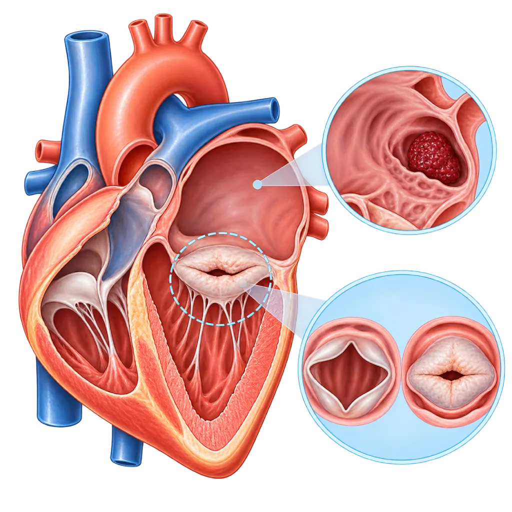
Mitral stenosis is a narrowing of the mitral valve, the doorway between the left atrium and the left ventricle of the heart. Blood is held back in the atrium and lungs, causing shortness of breath, palpitations, and tiring easily. The main cause is rheumatic heart disease.
SourcesSigns That Need Urgent Help
- Sudden severe shortness of breath, being unable to lie down, or noisy breathing
- Coughing up frothy or pink sputum
- Coughing up blood
- A drooping face, weakness in an arm or leg on one side, or slurred speech
- A very fast heartbeat with dizziness or severe shortness of breath
- Fainting
If these signs appear, go to the nearest emergency department right away or call Heart Attack Center 1500 258.
01 / Understanding the condition
About Mitral Stenosis
The mitral valve sits between the left atrium and the left ventricle of the heart. Each time the heart relaxes between two beats, this valve opens so that oxygen-rich blood from the lungs flows into the left ventricle. In mitral stenosis, the valve leaflets become thick and stiff and stick together at their edges, so the opening narrows. Blood is held back in the left atrium, the pressure in the lung blood vessels rises, and over time the left atrium enlarges. Mitral stenosis is one form of valve narrowing, covered on the Heart Valve Stenosis page.
Main Cause: Rheumatic Heart Disease
In Indonesia and other developing countries, the majority of mitral stenosis cases come from rheumatic fever, an inflammatory reaction after a strep throat infection that was not fully treated, usually in childhood and adolescence. This inflammation slowly damages the valve, and the narrowing often only becomes noticeable 10 to 20 years later, during the working-age years. In older adults, mitral stenosis can also be caused by calcification of the valve ring. Valve damage from rheumatic disease is covered on the Rheumatic Heart Disease page.
Symptoms That Develop Gradually
Symptoms usually appear slowly: shortness of breath during activity, tiring quickly, and palpitations. Many people unknowingly reduce their activity, so the symptoms seem not to get worse. Situations that make the heart beat faster, such as fever, pregnancy, anemia, or atrial fibrillation, can make shortness of breath suddenly worse, because the time to fill the left ventricle becomes shorter.
Atrial Fibrillation and Stroke Risk
An enlarged left atrium is prone to atrial fibrillation, an irregular heart rhythm. Blood that flows slowly in the atrium can form clots, which can break loose and block a blood vessel in the brain. That is why people with significant mitral stenosis and atrial fibrillation usually need an anticoagulant of the vitamin K antagonist type, monitored with blood tests, as decided by their doctor.
Treatment Can Be Done Through a Catheter
Medicines help reduce symptoms and prevent clots, but they do not widen the valve. When the narrowing is significant and causing symptoms, the valve can be widened with a balloon through a catheter (percutaneous mitral commissurotomy) if the valve shape is suitable. If it is not suitable, the valve is repaired or replaced through surgery.
02 / Understanding the condition
Symptoms to Recognize.
Common Symptoms
- Shortness of breath during activity, over time appearing with lighter and lighter activity
- Tiring easily and reduced ability to be active
- Palpitations or a heartbeat that feels irregular
- Shortness of breath when lying down, so you need to sleep on high pillows
- Coughing, especially at night or after activity
- Swelling in the ankles in the later stages
- Purplish-red cheeks in some people
Symptoms in Adult Women
Pregnancy increases blood volume and heart rate, so mitral stenosis is often first noticed during pregnancy.
- Shortness of breath that first appears or worsens during pregnancy, especially in the second and third trimesters
- A fast, pounding heartbeat during pregnancy
- Severe shortness of breath before, during, or after delivery
- Shortness of breath and tiredness mistaken for normal pregnancy complaints
03 / Understanding the condition
What Causes This Condition?
- Rheumatic fever in childhood or adolescence that slowly damages the valve (rheumatic heart disease)
- Calcification of the mitral valve ring and base in older age, often together with chronic kidney disease
- A congenital mitral valve abnormality, which is rare
- Other rare causes, such as radiation therapy to the chest or autoimmune diseases, for example lupus
04 / Understanding the condition
Who Is at Higher Risk?
Medical
A history of rheumatic fever, advanced chronic kidney disease, and radiation therapy to the chest area are linked to mitral valve damage.
Age
Rheumatic mitral stenosis often becomes noticeable between the ages of 20 and 50, while stenosis due to calcification generally appears in older adults.
Other
Repeated untreated sore throats, crowded housing, and limited access to health care in childhood increase the risk of rheumatic fever. Rheumatic mitral stenosis is also more common in women.
Groups at Higher Risk
- Rheumatic Fever Survivors: People who have had rheumatic fever, especially if they did not receive regular preventive antibiotics, are at risk of mitral valve damage years later.
- Women of Childbearing Age: Rheumatic mitral stenosis is more common in women and may first be noticed during pregnancy, so a heart checkup before pregnancy is recommended.
- Older Adults with Chronic Kidney Disease: Calcification of the mitral valve ring is more common in older adults and in people with advanced chronic kidney disease.
05 / Understanding the condition
How Is It Diagnosed?
Doctors usually suspect mitral stenosis from shortness of breath and a characteristic heart sound, a soft, low-pitched murmur over the apex of the heart. Echocardiography through the chest wall confirms the diagnosis. This test measures the area of the valve opening, the pressure difference across it, the pressure in the lung blood vessels, and the size of the left atrium, and it assesses the valve shape to decide whether balloon widening can be used.
Before balloon widening of the valve, echocardiography through the esophagus (transesophageal) is done to make sure there is no clot in the left atrium and to assess valve leaking. An ECG and heart rhythm recording look for atrial fibrillation. If symptoms and test results at rest do not match, echocardiography during activity helps assess the rise in lung pressure. Cardiac catheterization is used when other test results are still unclear.
Physical Examination
The doctor listens for the heart sound typical of mitral stenosis and looks for signs of fluid in the lungs or legs.
Echocardiography
The main test, including through the esophagus (TEE): measures the valve opening area, lung pressure, and left atrium size, and looks for clots before a procedure.
Electrocardiogram (ECG)
Looks for atrial fibrillation and signs of an enlarged left atrium.
Stress Echocardiography
Used when symptoms do not match the results at rest, to see the rise in pressure difference and lung pressure during activity.
Chest X-Ray
Shows enlargement of the left atrium and signs of fluid congestion in the lungs.
Cardiac Catheterization
Measures the pressure in the heart chambers directly when the results of tests without a catheter are still unclear.
06 / Understanding the condition
Treatment Options.
Treatment is decided by a doctor after an assessment. The categories below can help you follow the conversation with your doctor.
Medicines to Reduce Symptoms
Diuretics reduce excess fluid, while beta blockers or similar medicines slow the heart rate so the left ventricle has enough time to fill. Your doctor decides the type and dose.
Anticoagulants to Prevent Stroke
Given if there is atrial fibrillation, a history of clots, or a clot in the left atrium. In significant mitral stenosis, the main choice is a vitamin K antagonist monitored with the INR test.
Percutaneous Balloon Mitral Commissurotomy
A balloon is passed through a vein in the groin into the left atrium, then inflated to widen the valve without opening the chest. It is chosen when the valve shape is suitable and there is no clot or severe leaking.
Valve Repair or Replacement Surgery
Chosen when the valve is too stiff, calcified, or leaking for a balloon to be suitable, or when another valve problem needs surgery at the same time.
Secondary Prevention of Rheumatic Fever
Regular antibiotics, as injections or tablets on your doctor's schedule, to prevent rheumatic fever from coming back and the valve damage from getting worse.
07 / Understanding the condition
Complications to Watch For.
Atrial Fibrillation
An enlarged left atrium triggers an irregular rhythm that makes shortness of breath worsen quickly. Covered on the Atrial Fibrillation page.
Stroke and Embolism
Clots that form in the left atrium can break loose and block blood vessels in the brain or other organs. Covered on the Stroke page.
Pulmonary Hypertension
Long-lasting high pressure in the lung blood vessels strains the right ventricle of the heart. Covered on the Pulmonary Hypertension page.
Pulmonary Edema
Fluid builds up in the lungs when the heart beats fast, for example during pregnancy, fever, or atrial fibrillation, causing sudden severe shortness of breath. Covered on the Cardiogenic Pulmonary Edema page.
Right-Sided Heart Failure
When high lung pressure lasts a long time, the right ventricle weakens, leading to leg swelling, a swollen belly, and tricuspid valve leaking.
08 / Understanding the condition
What Can You Do?
- Take a child with a sore throat and fever to the doctor so that a bacterial infection is fully treated.
- If you have had rheumatic fever, take preventive antibiotics on your doctor's schedule without interruption.
- Have your heart checked before planning a pregnancy if you have a history of rheumatic fever or a heart murmur.
- Have regular echocardiography on schedule even if you have no symptoms yet.
- If you are already on a blood thinner, take it regularly and have your INR checked on schedule.
- Keep your teeth and mouth clean to reduce the risk of valve infection.
09 / Understanding the condition
When Should You See a Doctor?
See a cardiologist if you have a history of rheumatic fever, have been told you have a heart murmur, or start to feel short of breath and have palpitations during activity. A checkup before pregnancy is important for women with this history, because pregnancy adds to the heart's workload.
- Shortness of breath during light activity or when lying down
- Palpitations, or a heartbeat that feels irregular or very fast
- Coughing up blood or pink sputum
- Shortness of breath that first appears or worsens during pregnancy
- Signs of a stroke, such as a drooping face or weakness in one arm: go to the emergency room right away
Don't Wait in an Emergency.
If symptoms are severe or come on suddenly, go to the nearest emergency department right away or call Heart Attack Center 1500 258, rather than waiting for an appointment.
10 / Common questions
Questions You May Have.
Frequently asked questions about mitral stenosis.
Is mitral stenosis always caused by rheumatic heart disease?
Not always, but in Indonesia the majority of cases do come from rheumatic fever in childhood or adolescence. In older adults, especially those with chronic kidney disease, narrowing can occur because of calcification of the valve ring. Other causes are rare. Telling the causes apart matters, because rheumatic stenosis can often be widened with a balloon, while stenosis due to calcification is usually not suitable for a balloon.
Can women with mitral stenosis get pregnant?
Many women with mild mitral stenosis can go through pregnancy with close monitoring. In significant stenosis, pregnancy can trigger severe shortness of breath and fluid in the lungs, so doctors usually recommend widening the valve before pregnancy. Blood thinners also need special planning because some of them carry risks for the baby. Discuss pregnancy plans with a cardiologist and an obstetrician early on.
Does balloon widening of the valve require major surgery?
No. Percutaneous mitral commissurotomy is done through a catheter from a vein in the groin, without opening the chest. The catheter is guided to the left atrium, then a balloon is inflated to separate the edges of the valve leaflets that have stuck together. This procedure is suitable when the valve shape is right, there is no clot in the left atrium, and there is no severe leaking. If it is not suitable, your doctor will discuss surgical options.
Why is my blood thinner a warfarin-type medicine instead of a newer one?
In atrial fibrillation with significant mitral stenosis, guidelines recommend an anticoagulant of the vitamin K antagonist type, such as warfarin. A large study in people with rheumatic heart disease showed that this type was more effective at preventing stroke than one of the newer blood thinners. That is why regular INR tests are still needed. Do not switch or stop your medicine without your doctor's approval.
Can mitral stenosis come back after treatment?
Yes. After balloon widening, the valve can slowly narrow again over a few years to more than ten years, especially if rheumatic fever comes back. That is why regular checkups and echocardiography are still needed, and preventive antibiotics are continued on your doctor's schedule. If the valve narrows again, repeat widening or surgery can be considered depending on the valve shape at that time.
Sources and Medical Review
Translated from the Indonesian edition medically reviewed by Dr. dr. Dafsah A. Juzar, Sp.JP(K).
Sources as cited in the Indonesian edition.
- Vahanian A, Beyersdorf F, Praz F, Milojevic M, Baldus S, Bauersachs J, et al. 2021 ESC/EACTS Guidelines for the management of valvular heart disease. Eur Heart J. 2022;43(7):561-632.
- Praz F, et al. 2025 ESC/EACTS Guidelines for the management of valvular heart disease. Eur Heart J. 2025;46(44):4635-4736.
- Otto CM, Nishimura RA, Bonow RO, Carabello BA, Erwin JP 3rd, Gentile F, et al. 2020 ACC/AHA Guideline for the Management of Patients With Valvular Heart Disease: A Report of the American College of Cardiology/American Heart Association Joint Committee on Clinical Practice Guidelines. Circulation. 2021;143(5):e72-e227.
- Kumar RK, Antunes MJ, Beaton A, Mirabel M, Nkomo VT, Okello E, et al. Contemporary Diagnosis and Management of Rheumatic Heart Disease: Implications for Closing the Gap: A Scientific Statement From the American Heart Association. Circulation. 2020;142(20):e337-e357.
- Connolly SJ, Karthikeyan G, Ntsekhe M, Haileamlak A, El Sayed A, El Ghamrawy A, et al. Rivaroxaban in Rheumatic Heart Disease-Associated Atrial Fibrillation. N Engl J Med. 2022;387(11):978-988.
- Regitz-Zagrosek V, Roos-Hesselink JW, Bauersachs J, Blomström-Lundqvist C, Cífková R, De Bonis M, et al. 2018 ESC Guidelines for the management of cardiovascular diseases during pregnancy. Eur Heart J. 2018;39(34):3165-3241.
Doctors Who Treat This Condition

dr. Sri Diniharini, Sp.JP(K)
Echocardiography & Cardiovascular Imaging
Cardiovascular Diagnostic Center +1 center
Clinical focusAdvanced Echocardiography · Transesophageal Echocardiography
View Profile
Dr. dr. Dudy A. Hanafy, Sp.BTKV, Subsp. JD(K), MARS
Adult Cardiac Surgery
Advanced Cardiovascular Surgical Care +1 center
Clinical focusMinimally Invasive Cardiac Surgery · Coronary Artery Bypass Surgery
View Profile
Dr. dr. Dafsah A. Juzar, Sp.JP(K)
Interventional Cardiology
Coronary Intervention Procedures +1 center
Clinical focusComplex Coronary Intervention · Cardiac Catheterization
View Profile