
Quick answer
What Is Heart Valve Stenosis?
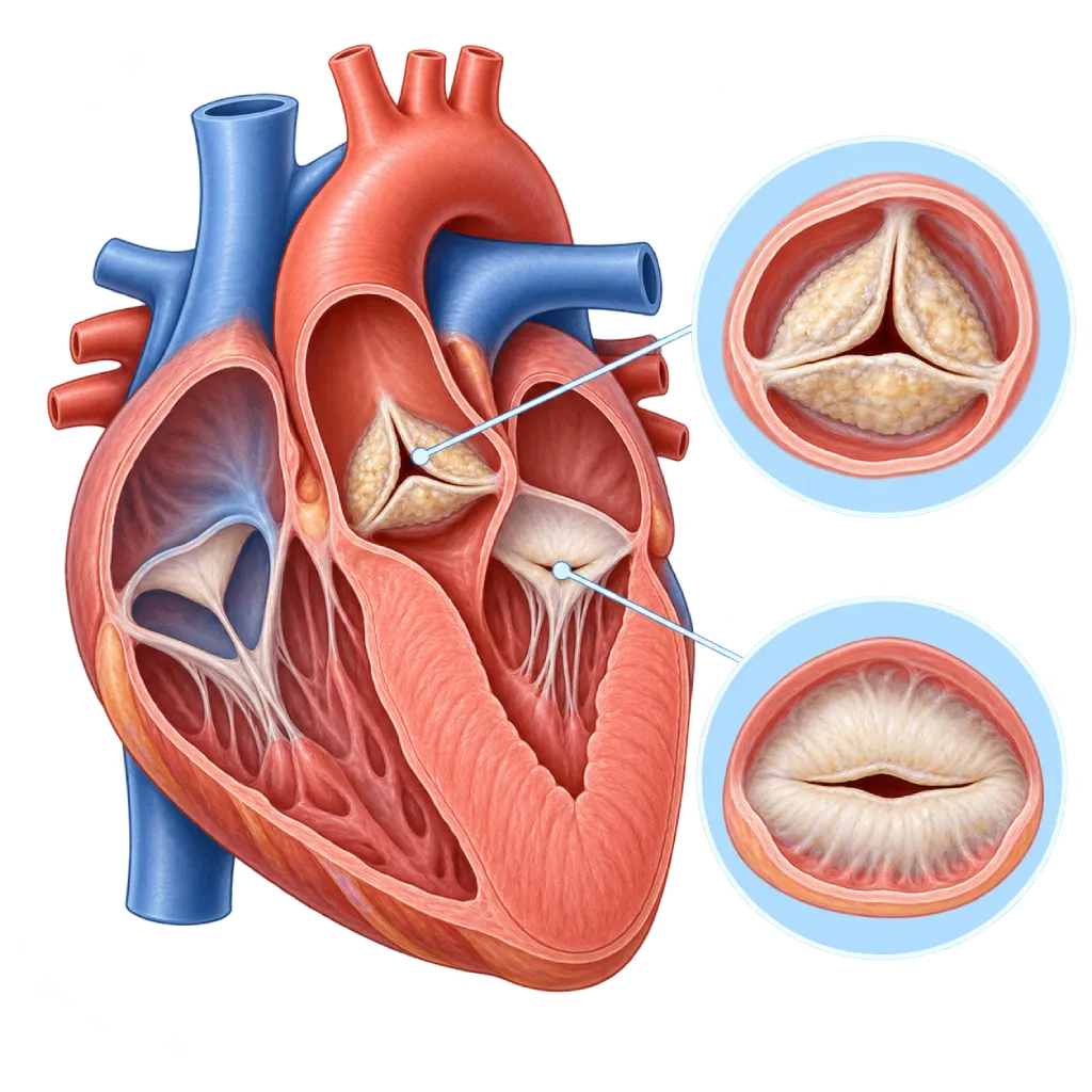
Heart valve stenosis is a narrowing of a heart valve, so the valve does not open wide and blood flow is obstructed. The heart has to pump harder, and symptoms usually develop slowly.
SourcesSigns That Need Urgent Help
- Fainting, especially during or shortly after activity
- Severe chest pain that does not ease with rest
- Severe shortness of breath at rest, or being unable to lie down
- Coughing up blood or pink, frothy sputum
- A very fast heartbeat with dizziness or shortness of breath
- Sudden weakness on one side of the body, facial drooping, or slurred speech
If these signs appear, go to the nearest emergency department right away or call Heart Attack Center 1500 258.
01 / Understanding the condition
About Heart Valve Stenosis
A healthy heart valve opens wide every time blood is pumped through it. In valve stenosis, the valve leaflets become thick, stiff, calcified, or stuck together, so the opening narrows. Blood has difficulty passing through, and the heart chamber behind the valve has to work harder.
Stenosis is different from regurgitation, in which the valve does not close tightly so blood flows backward. Both can occur together in one valve, for example in rheumatic heart disease, and they are often found together in more than one valve. Valve leakage is covered on the Valve Regurgitation / Insufficiency page.
Types of Valve Stenosis
Stenosis can affect all four valves, and each has its own page. Aortic stenosis, a narrowing at the outlet of the left ventricle, is common in older age. Mitral stenosis, a narrowing between the left atrium and the left ventricle, is usually caused by rheumatic heart disease. Pulmonary stenosis is usually a congenital condition present from birth. Tricuspid stenosis is rare and often accompanies other rheumatic valve disease.
What Happens to the Heart
In aortic stenosis, the left ventricle thickens to push blood through the narrow valve. Over time, the thickened muscle becomes stiff and does not get enough oxygen during activity, causing shortness of breath, chest pain, or dizziness. In mitral stenosis, pressure in the left atrium and the lung blood vessels rises, causing shortness of breath, and the atrium is prone to atrial fibrillation. In pulmonary stenosis, it is the right ventricle that bears the extra load.
Course of the Disease
The narrowing usually increases slowly over many years, and many people have no symptoms in the early stages. The 2021 ESC/EACTS and 2020 ACC/AHA guidelines emphasize two things: the degree of narrowing measured by echocardiography, and whether symptoms are present. Severe stenosis that already causes symptoms generally needs a procedure, because medication cannot widen the valve. In severe stenosis without symptoms, the decision is based on exercise test results, ventricular function, and other considerations.
Treatment options now vary, from widening the valve with a balloon and replacing the valve through a catheter to surgical valve replacement. The choice is tailored to the affected valve, the shape of the valve, your age, your general condition, and your preferences, after discussion with the heart team.
02 / Understanding the condition
Types and Related Conditions
The following conditions belong to the Heart Valve Stenosis group. Open each one for a full explanation.
03 / Understanding the condition
Symptoms to Recognize.
Common Symptoms
- Shortness of breath during activities that used to feel light
- Pain, heaviness, or pressure in the chest during activity
- Dizziness or near fainting during exercise or when climbing stairs
- Tiring quickly and a reduced ability to be active
- Palpitations or a heartbeat that feels irregular
- Coughing or shortness of breath when lying flat at night
- Swelling in the ankles or legs
Symptoms in Older Adults
Aortic stenosis due to calcification is common in older adults, and its symptoms are easily mistaken for normal signs of aging.
- Cutting back on activity without realizing it, to avoid shortness of breath
- Unsteadiness or near fainting when walking quickly
- Chest pain mistaken for a stomach or muscle problem
- Tiring quickly after everyday household chores
Symptoms in Adult Women
Rheumatic mitral stenosis often only becomes noticeable during pregnancy, because blood volume and heart rate increase.
- Shortness of breath that worsens in the second or third trimester
- Palpitations or an irregular rhythm that first appears during pregnancy
- Waking up at night because of shortness of breath or coughing
- Fatigue beyond the usual tiredness of pregnancy
Symptoms in Newborns
Very severe (critical) congenital pulmonary or aortic stenosis can cause symptoms in the first days or weeks of life.
- Bluish lips, skin, or nails
- Fast or labored breathing
- Poor feeding or tiring quickly while feeding
- Pale grayish skin that feels cold
- Difficulty gaining weight
04 / Understanding the condition
What Causes This Condition?
- Calcification and thickening of the aortic valve with age
- Rheumatic heart disease, which makes the mitral valve leaflets thicken and stick together
- A bicuspid (two-leaflet) aortic valve present from birth, which calcifies sooner
- Congenital pulmonary or aortic stenosis present from birth
- Calcification of the mitral valve ring in older adults or people with chronic kidney disease
- Late effects of radiation therapy to the chest area, for example in cancer treatment
- A biological artificial valve that wears out and narrows after many years
05 / Understanding the condition
Who Is at Higher Risk?
Age
The risk of aortic stenosis due to calcification rises markedly after age 65 to 70. With a bicuspid aortic valve, narrowing often appears one to two decades earlier.
Medical
A history of rheumatic fever, chronic kidney disease, high blood pressure, and chest radiation therapy increase the risk. Diabetes and high cholesterol are linked to aortic valve calcification.
Genetic
A bicuspid aortic valve can be found in several members of the same family. Inherited high levels of lipoprotein(a) are also linked to aortic valve calcification.
Lifestyle
Smoking is linked to aortic valve calcification, similar to the calcification process in blood vessels.
Other
Repeated untreated strep throat in childhood increases the risk of rheumatic fever, the main cause of mitral stenosis.
Groups at Higher Risk
- Older Adults: Aortic valve calcification becomes increasingly common after age 65, often without noticeable symptoms.
- Rheumatic Fever Survivors: Mitral valve narrowing can appear many years after rheumatic fever in childhood or adolescence.
- People with a Bicuspid Aortic Valve: A two-leaflet valve calcifies and narrows sooner, so regular echocardiograms are needed.
- People with Chronic Kidney Disease: Imbalances in calcium and phosphate can speed up calcification of the aortic valve and the mitral valve ring.
- Women with Mitral Stenosis Who Are Planning Pregnancy: Pregnancy adds to the heart's workload, so assessment and, if needed, a procedure should be done before pregnancy.
06 / Understanding the condition
How Is It Diagnosed?
Valve stenosis is often first suspected from a heart murmur that the doctor hears through a stethoscope. Echocardiography is the main test to see which valve is narrowed, measure the valve opening area and the speed of blood flow through it, and assess the thickness and strength of the heart chambers. From these results, the doctor determines the severity: mild, moderate, or severe.
If the results are unclear, especially when blood flow is low, the doctor may use stress echocardiography with a heart-stimulating medication, or a cardiac CT scan to measure the amount of calcium in the aortic valve. An exercise test helps confirm whether a person with severe stenosis truly has no symptoms. A CT scan is also used to plan transcatheter aortic valve implantation. Cardiac catheterization is done when the results do not agree, or to check the coronary arteries before a procedure.
Physical Examination
Listens for the heart murmur typical of valve narrowing, and checks the pulse, blood pressure, and signs of fluid buildup.
Echocardiography
The main test to measure the valve opening area, the speed and pressure difference of blood flow, and the thickness and function of the heart chambers.
Stress Echocardiography
Helps assess aortic stenosis with low blood flow, and assesses the rise in lung pressure during activity in mitral stenosis.
Cardiac CT Scan
Measures the amount of calcium in the aortic valve when the severity of stenosis is unclear, and maps the size of the valve and blood vessels for transcatheter valve implantation.
Treadmill Stress Test
An exercise test to confirm whether severe aortic stenosis is truly without symptoms; done only in people without symptoms, at the doctor's discretion.
Electrocardiogram (ECG)
Looks for thickening of the ventricular muscle, atrial fibrillation, and disorders of the heart's electrical conduction.
Cardiac Catheterization
Measures pressures directly when the results of other tests do not agree, and checks the coronary arteries before a valve procedure.
07 / Understanding the condition
Treatment Options.
Treatment is decided by a doctor after an assessment. The categories below can help you follow the conversation with your doctor.
Regular Monitoring
Mild to moderate stenosis without symptoms is monitored with regular checkups and echocardiograms; severe stenosis without symptoms is monitored more closely.
Medications to Relieve Symptoms
Diuretics, heart rate control medications, blood pressure medications, and blood thinners for atrial fibrillation help relieve symptoms but do not widen the valve. Your doctor determines the type.
Balloon Widening of the Valve
A balloon is inflated in the valve through a catheter to open leaflets that are stuck together, especially in rheumatic mitral stenosis with a suitable valve shape and in congenital pulmonary stenosis.
Transcatheter Aortic Valve Implantation
An artificial aortic valve is placed through a catheter from the groin without opening the chest, especially in older adults or patients with a higher surgical risk.
Valve Replacement Surgery
The narrowed valve is replaced with a mechanical or biological artificial valve through surgery, using a conventional incision or a small incision in selected patients.
08 / Understanding the condition
Complications to Watch For.
Heart Failure
A ventricle that keeps thickening or rising pressure in the lungs eventually causes shortness of breath, swelling, and fatigue. Covered on the Heart Failure page.
Atrial Fibrillation
An enlarged left atrium in mitral stenosis is prone to irregular rhythm. Covered on the Atrial Fibrillation page.
Stroke Caused by a Blood Clot
In mitral stenosis with atrial fibrillation, clots form easily in the left atrium and can travel to the brain.
Pulmonary Hypertension
Rising pressure in the left atrium is passed on to the lung blood vessels and strains the right ventricle. Covered on the Pulmonary Hypertension page.
Fainting and Dangerous Rhythm Disorders
In severe aortic stenosis, fainting during activity and ventricular rhythm disorders can occur, so these symptoms need to be checked right away.
Infective Endocarditis
A damaged valve is more easily infected by germs from the bloodstream. Covered on the Infective Endocarditis page.
09 / Understanding the condition
What Can You Do?
- Make sure sore throats in children are fully treated to prevent rheumatic fever
- If you have had rheumatic fever, follow long-term antibiotic prevention on the schedule your doctor sets
- Quit smoking and control your blood pressure, cholesterol, and blood sugar
- Keep your teeth and mouth clean to lower the risk of valve infection
- Have regular echocardiograms if you have a bicuspid aortic valve or a heart murmur
- Report new symptoms such as shortness of breath, chest pain, or dizziness during activity right away
- Plan pregnancy with a cardiologist if you have mitral or aortic stenosis
10 / Understanding the condition
When Should You See a Doctor?
See a cardiologist if a heart murmur is found or if you start to notice symptoms during activity. If you already know you have stenosis, the first appearance of symptoms is a reason to have a checkup sooner than scheduled, because this is often when a procedure needs to be considered. Go to the emergency room right away if you faint, have chest pain that does not ease, or have severe shortness of breath at rest.
- Shortness of breath, chest pain, or dizziness when walking quickly or climbing stairs
- A decline in your ability to be active compared with a few months earlier
- Palpitations with an irregular rhythm
- Being pregnant or planning a pregnancy with a history of valve disease
- A history of rheumatic fever or a bicuspid aortic valve without routine checkups
Don't Wait in an Emergency.
If symptoms are severe or come on suddenly, go to the nearest emergency department right away or call Heart Attack Center 1500 258, rather than waiting for an appointment.
11 / Common questions
Questions You May Have.
Frequently asked questions about heart valve stenosis.
Can valve stenosis be cured with medication?
Medication cannot widen a narrowed valve. It is used to relieve shortness of breath, regulate heart rate, control blood pressure, and prevent clots in atrial fibrillation. If the stenosis is severe and causes symptoms, the valve needs to be widened or replaced. In mild to moderate stenosis, regular monitoring is usually enough until your doctor determines that a procedure is needed.
What is the difference between balloon widening and valve replacement?
Balloon widening opens a valve whose leaflets are stuck together, and it is suitable for rheumatic mitral stenosis with certain valve shapes and for congenital pulmonary stenosis. In aortic stenosis caused by calcification, the results of a balloon alone do not last long, so the valve is generally replaced, either through surgery or through a catheter. Your doctor chooses based on the valve shape seen on echocardiography.
Is transcatheter aortic valve implantation suitable for everyone?
Not always. Valve implantation through a catheter is often chosen for older adults and patients with a higher surgical risk. For younger patients, certain bicuspid aortic valves, or people who also need other heart surgery, surgery may be more suitable. The heart team weighs your age, your anatomy on the CT scan, your general condition, and your preferences. Details are covered on the Transcatheter Aortic Valve Implantation (TAVI) page.
Can I exercise if I have valve stenosis?
Light to moderate activity is generally recommended in mild stenosis. In moderate or severe stenosis, especially aortic stenosis, strenuous or competitive sports and heavy lifting should be avoided until your doctor has assessed you. Stop the activity and get checked if you develop shortness of breath, chest pain, or dizziness during exercise. Your doctor may use an exercise test to determine safe limits.
Why does mitral stenosis often get worse during pregnancy?
During pregnancy, blood volume and heart rate increase. In mitral stenosis, the time for the left ventricle to fill becomes shorter, so pressure in the left atrium and lungs rises, causing shortness of breath. For this reason, women with moderate or severe mitral stenosis are advised to have an assessment before pregnancy and, if needed, to have the valve widened first.
How often do I need a repeat echocardiogram?
The interval depends on the severity and the type of valve. In mild stenosis, echocardiography is usually repeated every few years. In moderate stenosis, the interval is shorter, and severe stenosis without symptoms is generally monitored every 6 to 12 months. The test is done sooner if new symptoms appear. Your doctor will adjust the schedule to your condition.
Sources and Medical Review
Translated from the Indonesian edition medically reviewed by dr. BRM Ario S. Kuncoro, Sp.JP(K).
Sources as cited in the Indonesian edition.
- Vahanian A, Beyersdorf F, Praz F, Milojevic M, Baldus S, Bauersachs J, et al. 2021 ESC/EACTS Guidelines for the management of valvular heart disease. Eur Heart J. 2022;43(7):561-632.
- Otto CM, Nishimura RA, Bonow RO, Carabello BA, Erwin JP 3rd, Gentile F, et al. 2020 ACC/AHA Guideline for the Management of Patients With Valvular Heart Disease: A Report of the American College of Cardiology/American Heart Association Joint Committee on Clinical Practice Guidelines. Circulation. 2021;143(5):e72-e227.
- Baumgartner H, Hung J, Bermejo J, Chambers JB, Edvardsen T, Goldstein S, et al. Recommendations on the echocardiographic assessment of aortic valve stenosis: a focused update from the European Association of Cardiovascular Imaging and the American Society of Echocardiography. Eur Heart J Cardiovasc Imaging. 2017;18(3):254-275.
- Kumar RK, Antunes MJ, Beaton A, Mirabel M, Nkomo VT, Okello E, et al. Contemporary Diagnosis and Management of Rheumatic Heart Disease: Implications for Closing the Gap: A Scientific Statement From the American Heart Association. Circulation. 2020;142(20):e337-e357.
- Mack MJ, Leon MB, Thourani VH, Makkar R, Kodali SK, Russo M, et al. Transcatheter Aortic-Valve Replacement with a Balloon-Expandable Valve in Low-Risk Patients. N Engl J Med. 2019;380(18):1695-1705.
- Regitz-Zagrosek V, Roos-Hesselink JW, Bauersachs J, Blomstrom-Lundqvist C, Cifkova R, De Bonis M, et al. 2018 ESC Guidelines for the management of cardiovascular diseases during pregnancy. Eur Heart J. 2018;39(34):3165-3241.
- Baumgartner H, De Backer J, Babu-Narayan SV, Budts W, Chessa M, Diller GP, et al. 2020 ESC Guidelines for the management of adult congenital heart disease. Eur Heart J. 2021;42(6):563-645.
Doctors Who Treat This Condition

Dr. dr. Dafsah A. Juzar, Sp.JP(K)
Interventional Cardiology
Coronary Intervention Procedures +1 center
Clinical focusComplex Coronary Intervention · Cardiac Catheterization
View Profile
dr. BRM Ario S. Kuncoro, Sp.JP(K)
Echocardiography & Cardiovascular Imaging
Cardiovascular Diagnostic Center +2 centers
Clinical focusAdvanced and 3D Echocardiography · Transesophageal Echocardiography
View Profile
dr. Salomo Purba, Sp.BTKV, Subsp.JPK(K)
Pediatric and Congenital Heart Surgery
Congenital and Structural Heart Center +2 centers
Clinical focusPediatric Heart Surgery · Congenital Heart Defect Surgery
View Profile