
Quick answer
What Is Structural Heart Abnormalities?
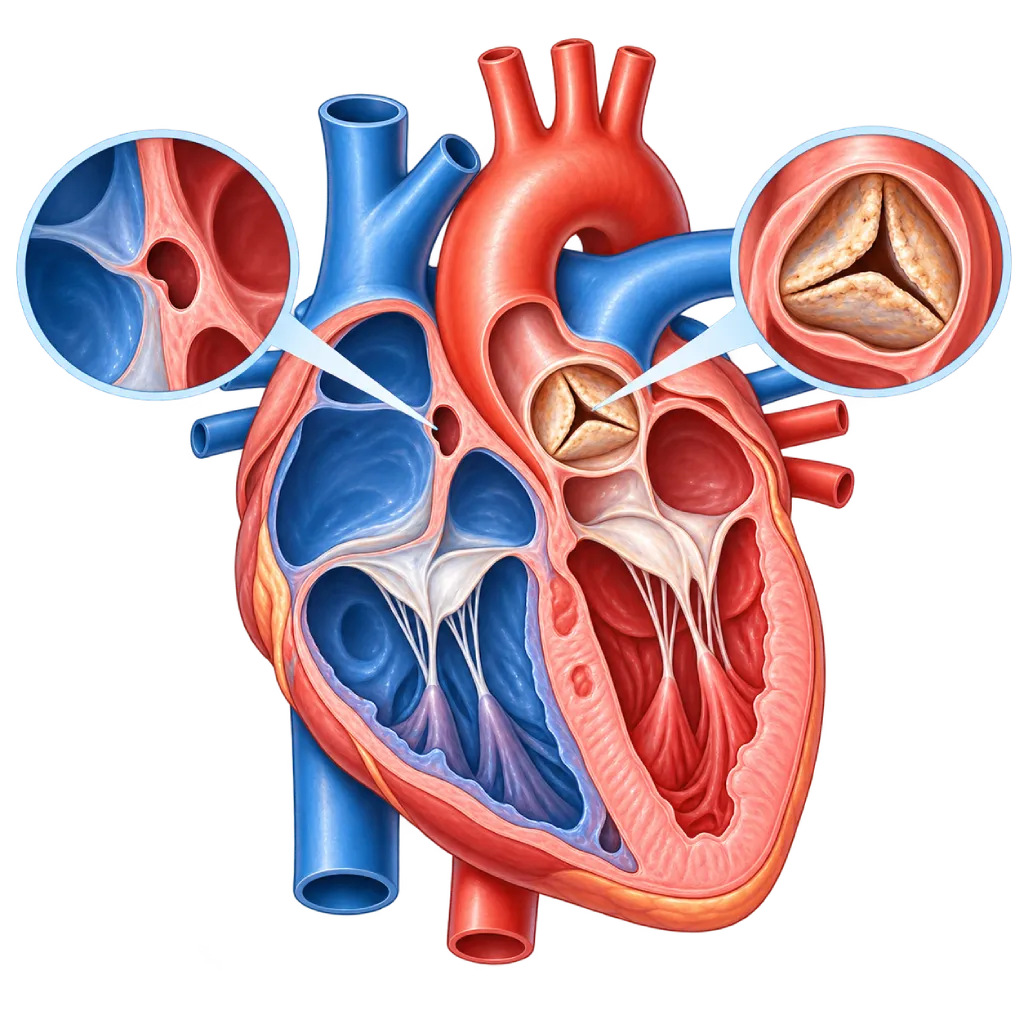
Structural heart abnormalities are changes in the physical parts of the heart, such as the dividing walls, valves, ventricle walls, or great vessels, that are present from birth or develop later. Their effects vary, from no symptoms to heart failure.
SourcesSigns That Need Urgent Help
- Severe shortness of breath at rest or a cough with frothy sputum
- Fainting, especially during activity
- Severe chest pain that starts suddenly
- A baby who looks very blue, limp, or hard to wake
- A very fast heartbeat with weakness or severe dizziness
If these signs appear, go to the nearest emergency department right away or call Heart Attack Center 1500 258.
01 / Understanding the condition
About Structural Heart Abnormalities
The heart is a muscular pump with four chambers. The two atria on top and the two ventricles below are separated by dividing walls (septa), four valves keep blood flowing in one direction, and great vessels such as the aorta and the pulmonary artery carry blood out of the heart. Structural heart abnormality is an umbrella term for changes in these physical parts. It is different from disease of the coronary arteries that supply the heart muscle, and from disorders of the electrical system that controls the heart rhythm.
Congenital or Acquired
Some abnormalities form while the fetal heart develops in the womb, for example a hole in a dividing wall, a narrowed valve, or an unusual position of the great vessels. This group is covered on the Congenital Heart Disease page. Others develop later, for example valves that calcify with age or are damaged by rheumatic fever, ventricle walls that thicken or enlarge because of heart muscle disease, and tears or bulges in the heart wall after a heart attack. Valve disorders are covered on the Heart Valve Disease page, while heart muscle disease is covered on the Cardiomyopathy page.
From Mild to Severe
Many structural abnormalities are mild. A small gap between the two atria, called a patent foramen ovale, is found in about one in four adults and generally does not need treatment. Small holes in the wall between a baby's ventricles often close on their own. In contrast, large abnormalities make the heart work harder or mix oxygen-rich blood with oxygen-poor blood, so they need early treatment.
How Abnormalities Are Found
Structural abnormalities are often first suspected from a heart murmur, an extra sound heard through a stethoscope, or from oxygen level screening in newborns. Echocardiography is the main test because it shows the shape and movement of each part of the heart without pain. In adults, congenital abnormalities are sometimes only discovered years later, and acquired abnormalities are found more often after the age of 65. Treatment options in adulthood, including catheter-based procedures, are covered on the Adult Structural Heart Disease page.
02 / Understanding the condition
Symptoms to Recognize.
Common Symptoms
Symptoms depend on the type and severity of the abnormality. Mild abnormalities often cause no symptoms.
- Shortness of breath during activity or when lying down
- Tiring quickly and a reduced ability to be physically active
- Palpitations or a heartbeat that feels irregular
- Swelling in the ankles or legs
- Chest pain or dizziness during activity
- No symptoms, with only a heart murmur found during an examination
Symptoms in Infants
Significant congenital abnormalities often show signs from infancy.
- Fast breathing or visible effort when breathing
- Tiring easily, sweating, or stopping during feeding
- Poor weight gain
- Bluish lips, tongue, or nails
- Frequent respiratory infections
Symptoms in Older Adults
In older age, common abnormalities are narrowing or leaking of the valves due to calcification.
- Shortness of breath or tiredness during activities that used to feel easy
- Dizziness or nearly fainting during activity
- Chest pain or heaviness when walking
- Swollen feet and difficulty sleeping while lying flat
03 / Understanding the condition
What Causes This Condition?
- Problems with the formation of the heart while the fetus develops in the womb
- Chromosomal or genetic abnormalities, for example Down syndrome
- Uncontrolled diabetes, rubella infection, or certain medications during pregnancy
- Calcification and aging of the valves over time
- Valve damage from rheumatic fever, covered on the Rheumatic Heart Disease page.
- Thickening or enlargement of the ventricle walls because of heart muscle disease or long-standing high blood pressure
- A tear in the dividing wall or a bulge in a ventricle wall after a heart attack
- Infection of a valve, covered on the Infective Endocarditis page.
04 / Understanding the condition
Who Is at Higher Risk?
Genetic
Congenital heart defects in a parent or sibling, chromosomal abnormalities, and inherited conditions such as Marfan syndrome and hypertrophic cardiomyopathy.
Medical
During pregnancy: uncontrolled diabetes and rubella infection. In adults: long-standing high blood pressure, heart attack, and chronic kidney disease.
Age
Congenital abnormalities usually appear in infancy, while valve calcification and changes in the heart walls become more common after the age of 65.
Other
A history of rheumatic fever in childhood, radiation therapy to the chest area, and exposure to alcohol, smoking, or certain medications while the fetus is developing.
Groups at Higher Risk
- Babies with Chromosomal Abnormalities: About half of babies with Down syndrome have a congenital heart defect, so they need echocardiography early on.
- Families of People with Congenital Heart Disease: Children of parents with a congenital heart defect have a higher risk of a similar defect.
- Rheumatic Fever Survivors: Rheumatic fever in childhood can damage the valves and only cause symptoms years later.
- Older Adults: Aortic valve calcification and mitral valve leakage are found more often with increasing age.
- Heart Attack Survivors: A large heart attack can change the shape of the left ventricle and cause mitral valve leakage.
05 / Understanding the condition
How Is It Diagnosed?
In babies, a severe structural abnormality may be suspected from oxygen level screening with a pulse oximeter before leaving the birthing facility, or from a heart murmur during a routine examination. In children and adults, the first clue is often a heart murmur, shortness of breath, or unusual findings on an ECG or chest X-ray.
Echocardiography is the next step because it shows the shape of the dividing walls, valves, and ventricle walls, as well as the direction of blood flow, without pain. If the images are not clear enough, the doctor may perform echocardiography through the esophagus. Cardiac MRI and CT give more detailed images of the anatomy, especially for complex congenital abnormalities or before a procedure. Cardiac catheterization is used when the pressures inside the heart and lungs need to be measured directly. From these results, the doctor determines the type of abnormality, its severity, and whether it can simply be monitored or needs treatment.
Physical Examination
The doctor listens to your heart with a stethoscope. A heart murmur, the pulse, and signs of fluid congestion are often the first clues.
Echocardiography
The main test to see the dividing walls, valves, ventricle walls, and blood flow. Echocardiography through the esophagus is used when more detailed images are needed.
Electrocardiogram (ECG)
Assesses the heart rhythm and signs of enlarged heart chambers that often accompany structural abnormalities.
Chest X-Ray
Shows the size of the heart and signs of excess blood flow to the lungs or fluid congestion.
Cardiac MRI
Measures chamber size, pumping strength, and blood flow in detail, especially in complex congenital abnormalities.
Cardiac CT Scan
Shows the anatomy of the great vessels, coronary arteries, and valves, for example to plan a procedure.
Cardiac Catheterization
Measures pressures and oxygen levels inside the heart chambers when other tests are not enough, sometimes as part of a procedure.
06 / Understanding the condition
Treatment Options.
Treatment is decided by a doctor after an assessment. The categories below can help you follow the conversation with your doctor.
Regular Monitoring
Mild abnormalities can simply be monitored with regular checkups and echocardiography to make sure they are not getting worse.
Medications to Control Symptoms
Diuretics, rhythm-control medications, or blood pressure-lowering medications reduce the strain on the heart and ease symptoms, but they do not correct the structure. Your doctor determines the type.
Catheter-Based Procedures
Many holes in the dividing walls and some valve abnormalities can now be closed or repaired through a blood vessel without opening the chest.
Heart Surgery
Chosen when the abnormality is complex, involves several parts of the heart, or is not suitable for catheter treatment.
Preventing Valve Infection
Keeping your teeth and mouth clean and having regular dental checkups. Antibiotics before certain dental procedures are given only to high-risk groups, based on your doctor's assessment.
Pregnancy Counseling
Women with a structural heart abnormality should have a consultation before pregnancy to assess the risks and prepare for monitoring.
07 / Understanding the condition
Complications to Watch For.
Heart Failure
A heart that keeps working harder because of a leak, a narrowing, or a large hole gradually weakens, causing shortness of breath and swelling.
Heart Rhythm Disorders
Enlarged heart chambers or scars from surgery can trigger irregular rhythms, for example atrial fibrillation.
Pulmonary Hypertension
Excess blood flow to the lungs from a large hole that has been left open for a long time can permanently raise the pressure in the lung blood vessels.
Infective Endocarditis
Abnormal valves or dividing walls are more easily infected by bacteria, so good dental and oral hygiene is important.
Stroke
Blood clots can form in an enlarged heart chamber or pass through a hole in a dividing wall, then travel to the brain.
Growth and Developmental Problems
In babies and children, severe abnormalities can hold back weight gain and development if left untreated.
08 / Understanding the condition
What Can You Do?
- Attend regular prenatal checkups and keep diabetes under control before and during pregnancy.
- Make sure your rubella immunization is complete before planning a pregnancy.
- Avoid smoking, alcohol, and medications without a prescription during pregnancy.
- Treat bacterial throat infections in children fully to prevent rheumatic fever.
- Control your blood pressure and heart attack risk factors in adulthood.
- Attend checkups and echocardiography as scheduled if you already know you have a structural abnormality.
- Keep your teeth and mouth clean to lower the risk of valve infection.
09 / Understanding the condition
When Should You See a Doctor?
Mild structural abnormalities often cause no symptoms, so a heart murmur or an unusual test result should be followed up with a cardiologist. If severe shortness of breath, fainting, or severe chest pain appears, or a baby looks very blue and limp, go to the nearest emergency room right away.
Make an appointment with a cardiologist if:
- A doctor hears a heart murmur in you or your child.
- Your baby tires easily while feeding, sweats a lot, or has poor weight gain.
- You are starting to get short of breath, tire quickly, or have palpitations during activities that used to be easy.
- You had heart surgery as a child and have not had a follow-up as an adult.
- You have a heart abnormality and are planning a pregnancy.
Don't Wait in an Emergency.
If symptoms are severe or come on suddenly, go to the nearest emergency department right away or call Heart Attack Center 1500 258, rather than waiting for an appointment.
10 / Common questions
Questions You May Have.
Frequently asked questions about structural heart abnormalities.
What is the difference between a structural heart abnormality and coronary artery disease?
Structural abnormalities involve the shape of the parts of the heart, such as the dividing walls, valves, and ventricle walls. Coronary artery disease involves the blood vessels that supply the heart muscle, covered on the Coronary Artery Disease page. Both can occur together, for example in an older adult with a narrowed aortic valve and narrowed coronary arteries, so doctors often check for both before deciding on a procedure.
Do all structural heart abnormalities need surgery?
No. Many mild abnormalities, such as a small gap in the wall between the atria or a mild valve leak, only need monitoring and do not interfere with daily life. A procedure is considered when the abnormality strains the heart, causes symptoms, or is at risk of getting worse. Many holes in the dividing walls and some valve abnormalities can now be treated through a catheter without opening the chest, so recovery is generally shorter.
My doctor heard a heart murmur. Does that mean there is a structural abnormality?
Not necessarily. Many children and some adults have a harmless heart murmur, which is heard because of fast blood flow through a normal heart, for example during a fever. However, a murmur can also come from a hole in a dividing wall or a valve that is narrowed or leaking. Echocardiography is a safe and painless way to confirm the cause.
Why can a congenital abnormality go undiscovered until adulthood?
Some abnormalities, for example a moderate hole in the wall between the atria or an aortic valve with only two leaflets, cause no symptoms for many years. The heart is able to adapt until the strain becomes too great, often in a person's 30s to 40s or during pregnancy. The first symptoms are usually shortness of breath, tiring quickly, or palpitations. Treatment options in adulthood are covered on the Adult Structural Heart Disease page.
Is pregnancy possible with a structural heart abnormality?
Many women with mild to moderate structural abnormalities can go through pregnancy safely when they are monitored. Pregnancy increases the heart's workload, so some severe abnormalities need to be treated before pregnancy, and a small number carry a high risk. Talk with a cardiologist before planning a pregnancy so the risks can be assessed and monitoring during pregnancy and delivery can be prepared.
What is meant by a "leaky heart"?
"Leaky heart" is a lay term usually used for a hole in the dividing wall of the heart (often called a hole in the heart), for example an atrial septal defect or a ventricular septal defect. Sometimes the term is also used for a valve that does not close tightly. Both are structural abnormalities, but their treatment differs. So ask your doctor which part is meant, how large the problem is, and whether it needs to be closed or can simply be monitored.
Sources and Medical Review
Translated from the Indonesian edition medically reviewed by dr. BRM Ario S. Kuncoro, Sp.JP(K).
Sources as cited in the Indonesian edition.
- Baumgartner H, De Backer J, Babu-Narayan SV, Budts W, Chessa M, Diller GP, et al. 2020 ESC Guidelines for the management of adult congenital heart disease. Eur Heart J. 2021;42(6):563-645.
- Praz F, Borger MA, Lanz J, Marin-Cuartas M, Abreu A, Adamo M, et al. 2025 ESC/EACTS Guidelines for the management of valvular heart disease. Eur Heart J. 2025;46(44):4635-736.
- Arbelo E, Protonotarios A, Gimeno JR, Arbustini E, Barriales-Villa R, Basso C, et al. 2023 ESC Guidelines for the management of cardiomyopathies. Eur Heart J. 2023;44(37):3503-626.
- Liu Y, Chen S, Zuhlke L, Black GC, Choy MK, Li N, et al. Global birth prevalence of congenital heart defects 1970-2017: updated systematic review and meta-analysis of 260 studies. Int J Epidemiol. 2019;48(2):455-63.
- Pierpont ME, Brueckner M, Chung WK, Garg V, Lacro RV, McGuire AL, et al. Genetic Basis for Congenital Heart Disease: Revisited: A Scientific Statement From the American Heart Association. Circulation. 2018;138(21):e653-711.
- Martin GR, Ewer AK, Gaviglio A, Hom LA, Saarinen A, Sontag M, et al. Updated Strategies for Pulse Oximetry Screening for Critical Congenital Heart Disease. Pediatrics. 2020;146(1):e20191650.
- De Backer J, Haugaa KH, Hasselberg NE, de Hosson M, Brida M, Castelletti S, et al. 2025 ESC Guidelines for the management of cardiovascular disease and pregnancy. Eur Heart J. 2025;46(43):4462-568.
Doctors Who Treat This Condition

dr. BRM Ario S. Kuncoro, Sp.JP(K)
Echocardiography & Cardiovascular Imaging
Cardiovascular Diagnostic Center +2 centers
Clinical focusAdvanced and 3D Echocardiography · Transesophageal Echocardiography
View Profile
dr. Radityo Prakoso, Sp.JP(K)
Pediatrics & Congenital Heart Disease
Congenital and Structural Heart Center
Clinical focusPediatric Cardiology Consultation · Congenital Heart Disease Care
View Profile
dr. Salomo Purba, Sp.BTKV, Subsp.JPK(K)
Pediatric and Congenital Heart Surgery
Congenital and Structural Heart Center +2 centers
Clinical focusPediatric Heart Surgery · Congenital Heart Defect Surgery
View Profile