
Quick answer
What Is Heart Defect Closure?
Heart defect closure is a procedure to close a congenital hole in the heart's septum (dividing wall) or the channel between the aorta and pulmonary artery, such as ASD, VSD, and PDA. It stops the blood leak that strains the heart and lungs. Many holes can be closed through a catheter in 1 to 2 hours; others need surgery.
Sources01 / Understanding the procedure
About Heart Defect Closure
What Is Heart Defect Closure
A heart defect is a hole or channel in the heart that should not be there, usually present from birth. This hole lets some oxygen-rich blood leak back to the right side of the heart and into the lungs, so the heart and lungs have to work harder. Heart defect closure is a procedure to close the hole, either through a catheter or through surgery.
Defects That Are Commonly Closed
- ASD (atrial septal defect): a hole in the wall between the two upper chambers of the heart (atria). This is covered on the Atrial Septal Defect (ASD) Closure page.
- VSD (ventricular septal defect): a hole in the wall between the two lower chambers of the heart (ventricles). This is covered on the Ventricular Septal Defect (VSD) Closure page.
- PDA (patent ductus arteriosus): a channel between the aorta and the pulmonary artery that should close after birth. This is covered on the Patent Ductus Arteriosus (PDA) Closure page.
- PFO (patent foramen ovale): a small flap-like opening in the wall between the upper chambers, found in about one in four adults. It usually does not need to be closed, except in certain situations such as a stroke with no other clear cause. This is covered on the Patent Foramen Ovale page.
Catheter or Surgery
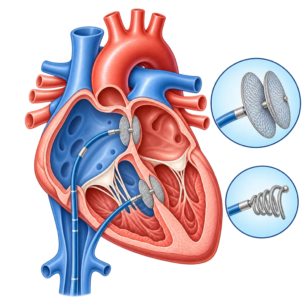
In catheter closure, the doctor inserts a thin tube through a blood vessel in the groin up to the heart, then places a closure device. The device is usually made of two flexible metal mesh discs that clamp the edges of the hole, or a coil for small channels. Within a few months, heart tissue grows over the device. This approach does not require a cut in the chest, and recovery is short.
Surgery is chosen when the hole is too large, the rim of tissue around it is too small, it lies close to a valve or the heart's electrical pathway, or another abnormality needs to be repaired at the same time. The hole is closed with stitches or a patch, usually with the help of a heart-lung machine; this surgery is covered on the Congenital Heart Surgery page.
When a Hole Needs to Be Closed
Not every hole needs to be closed. Small holes, especially in babies, often get smaller or close on their own. Closure is recommended when the leak is large enough to enlarge the heart chambers, the baby has trouble feeding and growing, shortness of breath develops, or there has been an infection of the heart's inner lining. If left untreated, a large leak can cause high pressure in the lung blood vessels, heart rhythm disorders, and heart failure.
Tests That Guide the Choice
Echocardiography is the main test to see the type, size, and rim of the hole. In adults, an echocardiogram through the esophagus (TEE) is often needed for a clearer picture. If the lung pressure is suspected to be high, the doctor measures it directly with right heart catheterization, covered on the Right Heart Catheterization page. Lung pressure that is already very high can make closure no longer safe.
Children and Adults
In children, the timing of closure is adjusted to the size of the hole and the child's growth, and parents are involved in every decision. Some people only find out about their hole as adults, for example when shortness of breath or palpitations appear. Closure is still beneficial in adulthood as long as the lung pressure is not yet too high.
02 / Understanding the procedure
When Is This Procedure Considered?
A doctor may consider this procedure in the situations below. The decision is always made after a thorough assessment.
- A Leak That Strains the HeartThe heart chambers become enlarged because of excess blood flow through the hole or channel.
- Baby Has Trouble Feeding and GrowingThe baby sweats and tires quickly while feeding, breathes fast, and has trouble gaining weight.
- Shortness of Breath or Tiring EasilySymptoms during activity in older children or adults that are related to the leak.
- History of EndocarditisA past infection of the heart's inner lining caused by the forceful blood flow through the hole or channel.
- Stroke with a Patent Foramen OvaleA stroke with no other clear cause, especially under the age of 60, after assessment together with a neurologist.
Situations to Consider First
- Lung Pressure Already Very HighWhen blood has already reversed to flow from right to left (Eisenmenger syndrome), closure would actually be harmful.
- Small Hole Without Strain on the HeartA small hole that does not strain the heart only needs regular monitoring.
- Anatomy Not Suitable for a CatheterThe hole is too large, lacks enough rim, or is close to a valve; surgery becomes the option.
- Active Infection or a Clot in the HeartA planned procedure is postponed until the infection is resolved or the clot is gone.
03 / Before the procedure
How Should You Prepare?
Preparation depends on the type of hole, age, and closure method. The team will give you written instructions.
Tests before the procedure. Usually echocardiography, ECG, chest X-ray, and blood tests. In adults, an echocardiogram through the esophagus (TEE) is often added.
Fasting. Follow the fasting schedule from the anesthesia team. For babies and children, the time limits for breast milk, formula, and water are explained separately.
Medicines and allergies. Bring a list of your medicines and supplements. Tell the team about any allergy to medicines, contrast dye, or metals such as nickel.
Dental health and infections. Have your teeth checked before a planned procedure. Tell the team if there is a fever, cough, or cold.
Preparing your child. Explain what will happen in simple words. Parents can stay with their child until the anesthesia takes effect, and the child may bring a favorite item.
Unsure about the preparation instructions? Ask our team before the procedure.
04 / During the procedure
How Is the Procedure Done?
Catheter closure is done in the catheterization lab and takes about 1 to 2 hours. Children usually receive general anesthesia; adults may receive general anesthesia or a sedative with local anesthesia. Surgery is done in the operating room under general anesthesia and takes longer, about 3 to 5 hours.
Inserting the Catheter
The doctor inserts a thin tube through a blood vessel in the groin. A blood thinner is given so that clots do not form during the procedure.
Measuring the Hole
The pressures in the heart and lungs are measured, then the hole is viewed with echocardiography and X-ray to choose the right device size.
Placing the Closure Device
The device, folded inside the catheter, is pushed to the hole and then opened so that it clamps both sides of the hole's edge. Before the device is released, its position and seal are checked.
Completing the Procedure
The catheter is removed and the puncture site is pressed until the bleeding stops. The patient is then monitored on the ward.
05 / Benefits and risks
Benefits and Risks Should Be Discussed Together.
Every medical procedure has benefits and risks. Your doctor will explain both for your situation before the procedure.
Benefits You Can Expect
- Less Strain on the Heart and LungsThe leak stops, so enlarged heart chambers can shrink back.
- No Cut in the ChestIn catheter closure, the device is inserted from the groin, and patients usually go home within 1 to 2 days.
- Better Growth in ChildrenBabies with a large leak usually feed more easily and gain weight after the hole is closed.
- Preventing Long-Term ComplicationsLowers the risk of high lung pressure, heart rhythm disorders, and heart failure later in life.
The benefits of a procedure are not a promise of a cure. Ask how the results will affect your care plan.
Risks You Should Know
- Device Moving Out of PlaceRare; the device can usually be retrieved through a catheter, and occasionally surgery is needed.
- Heart Rhythm DisordersAn irregular or slowed heartbeat, mostly temporary; a pacemaker is rarely needed.
- Problems at the Puncture SiteBruising, bleeding, or reduced blood flow to the leg, more likely in babies.
- Residual LeakA small leak may remain at the edge of the device or patch and often gets smaller over time.
- Risks with SurgeryBleeding, wound infection, and fluid around the heart, which are monitored in the intensive care unit.
Risks can differ from person to person. A full explanation and consent are given after an individual assessment.
06 / After the procedure
What Is Recovery Like?
After catheter closure, most patients go home within 1 to 2 days. After surgery, the hospital stay is usually about one week, including a few days in the intensive care unit.
First day. Lie with your legs straight for a few hours. An echocardiogram is done before going home to confirm the device position.
Medicines. After a device is placed in the heart wall, the doctor usually prescribes an antiplatelet medicine such as aspirin for about 6 months.
Activity. Light activity and school can resume within a few days. Strenuous exercise and contact sports are postponed for a few weeks as advised.
Dental health. For 6 months after a device or patch is placed, tell your dentist before any dental procedure, because preventive antibiotics may be needed.
Follow-up visits. Echocardiography is done regularly, for example at 1, 6, and 12 months, then as advised.
Warning signs. Go to the emergency room right away if you or your child has:
- Chest pain, shortness of breath, or fainting
- A very fast or irregular heartbeat
- Growing swelling or bleeding at the puncture site
- High fever with no clear cause
- Facial drooping, weakness on one side of the body, or slurred speech
Know the Signs That Need Prompt Care.
Follow the team's guidance on warning signs. If severe or sudden symptoms appear after the procedure, go to the nearest emergency department right away or call Heart Attack Center 1500 258.
07 / Team and support
Doctors and Procedure Support
The doctors below are listed for this procedure. Listed by name, not by ranking.

dr. Radityo Prakoso, Sp.JP(K)
Pediatrics & Congenital Heart Disease
Congenital and Structural Heart Center
Clinical focusPediatric Cardiology Consultation · Congenital Heart Disease Care
View Profile
dr. Salomo Purba, Sp.BTKV, Subsp.JPK(K)
Pediatric and Congenital Heart Surgery
Congenital and Structural Heart Center +2 centers
Clinical focusPediatric Heart Surgery · Congenital Heart Defect Surgery
View ProfileCatheterization Laboratory (Cath Lab)
A dedicated procedure room for cardiac catheterization, coronary angiography, stent placement, and other catheter-based heart…
The availability of technology and test support is confirmed with the team. Technology is chosen according to clinical need.
08 / Common questions
Before You Plan the Procedure.
Questions patients and parents often ask about heart defect closure.
Can the hole in my child's heart close on its own?
Yes, especially small holes. Many small VSDs and some small ASDs get smaller or close on their own in the first years of life. A PDA in a full-term baby that is still open after a few months rarely closes on its own. The doctor will monitor it with regular echocardiograms and recommend closure if the hole strains the heart or shows no sign of getting smaller.
Which is better, catheter closure or surgery?
Both aim to close the hole. Catheter closure needs no cut in the chest and has a shorter recovery, but it only suits holes of a certain size, location, and rim. Surgery can close almost any type of hole and repair other abnormalities at the same time. The heart team will recommend the suitable approach based on the echocardiogram results and the patient's condition.
Will the closure device stay in the heart?
Yes. The device is left in place and does not need to be replaced. Within a few months, heart tissue grows over it, so the device becomes part of the heart wall. Keep the device card you receive after the procedure, and show it to health workers before tests such as an MRI, because some devices require special settings.
Is closing a hole in the heart painful?
During the procedure, the patient is given anesthesia and does not feel pain. After catheter closure, there is usually only some soreness or bruising in the groin for a few days. After surgery, pain at the chest wound is treated with pain relievers and gradually eases over a few weeks. Some people feel brief palpitations in the first few weeks.
Do adults still need to have a hole in the heart closed?
Yes, if the hole still strains the heart, for example when the right heart chambers are enlarged, even if there are no symptoms yet. Closure in adulthood can relieve shortness of breath and improve exercise tolerance. The condition is that the pressure in the lung blood vessels is not yet too high. For this reason, the doctor may perform right heart catheterization to measure it before deciding.
What is a PFO, and does it have to be closed?
A PFO is a small opening in the wall between the upper heart chambers that should close after birth but stays open in about one in four adults. The majority cause no problems and do not need to be closed. Closure is considered for people who have had a stroke with no other clear cause, after assessment by both a cardiologist and a neurologist.
Sources and Medical Review
Translated from the Indonesian edition medically reviewed by dr. Radityo Prakoso, Sp.JP(K).
Sources as cited in the Indonesian edition.
- Baumgartner H, De Backer J, Babu-Narayan SV, et al. 2020 ESC Guidelines for the management of adult congenital heart disease. Eur Heart J. 2021;42(6):563-645.
- Stout KK, Daniels CJ, Aboulhosn JA, et al. 2018 AHA/ACC Guideline for the Management of Adults With Congenital Heart Disease: A Report of the American College of Cardiology/American Heart Association Task Force on Clinical Practice Guidelines. Circulation. 2019;139(14):e698-e800.
- Feltes TF, Bacha E, Beekman RH, et al. Indications for Cardiac Catheterization and Intervention in Pediatric Cardiac Disease: A Scientific Statement From the American Heart Association. Circulation. 2011;123(22):2607-2652.
- Pristipino C, Sievert H, D'Ascenzo F, et al. European position paper on the management of patients with patent foramen ovale. General approach and left circulation thromboembolism. Eur Heart J. 2019;40(38):3182-3195.
- Delgado V, Ajmone Marsan N, de Waha S, et al. 2023 ESC Guidelines for the management of endocarditis. Eur Heart J. 2023;44(39):3948-4042.