
Quick answer
What Is Ventricular Septal Defect?
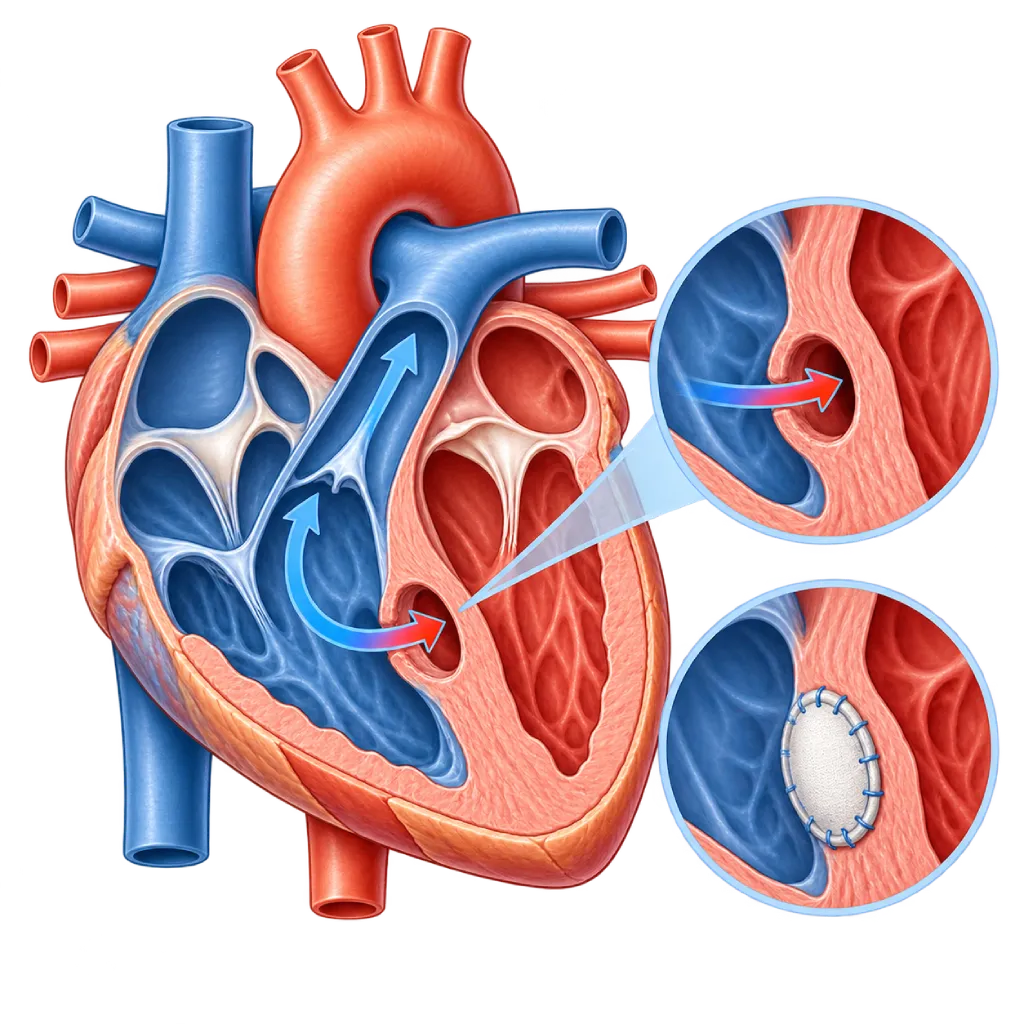
A ventricular septal defect is a hole, present from birth, in the wall between the two lower chambers of the heart. Small holes often close on their own, while large holes can cause heart failure in babies and need to be closed.
SourcesSigns That Need Urgent Help
- Lips, tongue, or nails that look bluish
- Very fast breathing, gasping, or the chest pulling inward
- A baby refusing to feed or looking very limp
- Fainting, or a very fast heartbeat that does not settle
- Pale grayish skin with cold hands and feet
If these signs appear, go to the nearest emergency department right away or call Heart Attack Center 1500 258.
01 / Understanding the condition
About Ventricular Septal Defect
The heart has two lower chambers (ventricles) separated by a thick wall called the ventricular septum. In a ventricular septal defect (VSD), this wall has had a hole since birth. The pressure in the left ventricle is much higher than in the right ventricle, so oxygen-rich blood is pushed from left to right and back to the lungs. As a result, the lungs receive too much blood and the left side of the heart has to work harder. VSD is a common congenital heart defect in children. It can occur on its own or as part of another defect, such as tetralogy of Fallot.
Types and Size of the Hole
By location, a perimembranous VSD lies near the valves, a muscular VSD lies in the muscular part of the septum, and a subarterial VSD lies just below the aortic valve and the pulmonary valve. Subarterial VSD is found more often in children in Asia and can cause a leaflet of the aortic valve to sag and then leak. However, the size of the hole matters more for its effects. A small hole lets only a little blood through and often causes a loud heart murmur without symptoms. A large hole causes signs of heart failure in the first months of life. A moderate hole falls between the two and needs closer monitoring.
Course of the Condition
Many small VSDs, especially the muscular type, close on their own in the first years of life. With a large hole that is not closed, the excess flow gradually damages the lung blood vessels. If the pressure in the lungs becomes very high, the flow can reverse from right to left and the body turns bluish. This is called Eisenmenger syndrome, and it can no longer be corrected by closing the hole. For this reason, large holes are usually closed in the first year of life.
In Adulthood
Adults with a small VSD that does not strain the heart can usually live normal lives, but they still need checkups because of the risk of endocarditis and aortic valve leakage. The 2020 ESC and 2018 AHA/ACC guidelines recommend closure in adults when the left ventricle is enlarged because of the shunt flow and the pressure in the lungs is not yet too high. Adults who had surgery as children are also advised to have regular checkups to assess any residual hole, the valves, and the heart rhythm.
02 / Understanding the condition
Symptoms to Recognize.
Common Symptoms
- A loud heart murmur on examination, often without other complaints
- Tiring quickly or getting short of breath during activity with a moderate to large hole
- Frequent coughs and colds or repeated pneumonia
- Palpitations or a pulse that feels irregular in adults
Symptoms in Infants
With a large hole, symptoms usually start to appear a few weeks after birth, often at 1 to 2 months of age.
- Fast breathing, especially during feeding
- Heavy sweating on the head during feeding
- Often stopping feeding because of tiredness, so feeds take a long time
- Difficulty gaining weight
- Fussiness, pale skin, and tiring easily
Symptoms in Children
- Growth that lags behind other children of the same age
- Tiring more quickly during play
- Frequent pneumonia or respiratory infections
03 / Understanding the condition
What Causes This Condition?
- The wall between the two lower chambers does not close completely while the heart is forming in the womb
- In most children, the exact trigger is unknown
- Chromosomal disorders, such as Down syndrome, trisomy 13, and trisomy 18
- Certain gene changes that can run in families
- Pregnancy factors, such as uncontrolled diabetes, rubella infection, alcohol, and certain medicines
04 / Understanding the condition
Who Is at Higher Risk?
Genetic
A history of congenital heart disease in a parent or sibling and genetic syndromes such as Down syndrome increase the likelihood of VSD.
Medical
Uncontrolled diabetes, obesity, and rubella infection in the mother in early pregnancy are linked to a higher risk of heart defects in the baby.
Lifestyle
Smoking, drinking alcohol, and using medicines without a prescription during pregnancy are linked to a higher risk of birth defects, including septal defects of the heart.
Other
Premature babies and babies with other birth defects more often have a VSD.
Groups at Higher Risk
- Babies with a Large Hole: At risk of heart failure and poor growth in the first months, so they need close monitoring and timely closure.
- Children with Down Syndrome: Heart defects often accompany Down syndrome, including VSD and atrioventricular septal defect, so echocardiography is recommended after birth.
- People with Subarterial VSD: A hole just below the aortic valve can make the valve sag and leak even when the hole is small, so it often needs to be closed earlier.
- Adults with an Unclosed VSD: Still at risk of endocarditis, aortic valve leakage, and rhythm problems, so regular checkups remain necessary.
05 / Understanding the condition
How Is It Diagnosed?
Many VSDs are found because a doctor hears a heart murmur when examining a baby, often at a few weeks of age. A small hole can actually cause a loud murmur. In a baby with fast breathing and difficulty gaining weight, the doctor also looks for signs of heart failure, such as an enlarged liver.
Echocardiography confirms the diagnosis. This test shows the location and size of the hole, the direction of blood flow, the size of the left ventricle, the estimated pressure in the lungs, and whether the aortic valve is also affected. An ECG and a chest X-ray complete the assessment of the strain on the heart. Cardiac catheterization is done when the pressure in the lungs is suspected to be high or when the hole is to be closed through a catheter. In teenagers and adults, cardiac MRI can help measure the size of the shunt.
Physical Examination
The doctor hears a typical heart murmur along the left side of the breastbone. A small hole often actually sounds louder.
Echocardiography
The main test to determine the location and size of the hole, the direction of flow, the strain on the left ventricle, the estimated lung pressure, and the condition of the aortic valve.
Electrocardiogram (ECG)
Shows signs of strain on the left or right ventricle and rhythm problems.
Chest X-Ray
Shows heart enlargement and excess blood flow to the lungs.
Cardiac Catheterization
Measures the pressure and resistance in the lung blood vessels when they are suspected to be high, to make sure the hole is still safe to close.
Cardiac MRI
In teenagers and adults, measures the size of the left ventricle and the amount of shunt flow when echocardiography results are unclear.
06 / Understanding the condition
Treatment Options.
Treatment is decided by a doctor after an assessment. The categories below can help you follow the conversation with your doctor.
Regular Monitoring
A small VSD that does not strain the heart and does not cause aortic valve leakage only needs monitoring. Many of these close on their own.
Heart Failure Medicines for Babies
Diuretics and other medicines can ease breathlessness while waiting for the hole to get smaller or be closed. The type and dose are decided by the doctor.
Nutritional Support
Babies with a large hole need more calories, for example fortified breast milk or formula and a planned feeding schedule, as directed by the doctor.
Surgical Closure
The hole is closed with a patch during open-heart surgery. This approach is used for the majority of large VSDs in babies. Covered on the Congenital Heart Surgery page.
Catheter-Based Closure
Muscular VSDs and some perimembranous VSDs in older children or adults can be closed with a device through a catheter, after a careful assessment of the anatomy.
Long-Term Follow-Up
After closure, regular checkups assess any residual hole, the aortic valve, the heart rhythm, and the pressure in the lungs.
07 / Understanding the condition
Complications to Watch For.
Heart Failure in Babies
A large hole makes the lungs receive too much blood, so the baby becomes short of breath, sweats, and has difficulty gaining weight.
Pulmonary Hypertension and Eisenmenger Syndrome
Years of excess flow damage the lung blood vessels. If the flow reverses, the body turns bluish and the hole can no longer be closed. Covered on the Pulmonary Hypertension page.
Aortic Valve Leakage
In a subarterial or perimembranous VSD, a leaflet of the aortic valve can sag into the hole and leak, which is often a reason for earlier closure.
Infective Endocarditis
The strong jet of blood through the hole can injure the inner lining of the heart, making it easier for germs to attach. Covered on the Infective Endocarditis page.
Rhythm Problems and Heart Block
Rhythm problems can occur in adults. Heart block can be a rare complication after closure, especially in perimembranous VSD.
08 / Understanding the condition
What Can You Do?
- Have regular prenatal checkups, including an ultrasound that assesses the baby's heart
- Make sure you are immune to rubella before pregnancy
- Control your blood sugar before and during pregnancy
- Avoid smoking, alcohol, and medicines without a prescription during pregnancy
- For people with a VSD, brush your teeth regularly and have a dental checkup every 6 months
- Keep to the checkup schedule even if your child seems healthy
09 / Understanding the condition
When Should You See a Doctor?
Take your baby or child to a doctor if the following signs appear. Go to the emergency room right away if your baby looks bluish, is breathing very hard, or refuses to feed.
- Your baby breathes fast or sweats during feeding
- Your baby often stops feeding and has difficulty gaining weight
- A doctor hears a heart murmur during an immunization visit or a routine checkup
- Your child often gets pneumonia or tires quickly during play
- A person with a VSD has a fever lasting more than a few days without a clear cause
- An adult with a VSD has palpitations, shortness of breath, or tires quickly
Don't Wait in an Emergency.
If symptoms are severe or come on suddenly, go to the nearest emergency department right away or call Heart Attack Center 1500 258, rather than waiting for an appointment.
10 / Common questions
Questions You May Have.
Questions parents often ask about ventricular septal defect.
Is a small VSD dangerous?
Usually not. A small VSD often sounds like a loud murmur, but only a little blood flows through it and it does not strain the heart. Many close on their own, especially the muscular type. Children with a small VSD can grow and be active like other children. Checkups are still needed because there is a small risk of endocarditis and, in certain locations, aortic valve leakage.
When does a VSD need to be closed?
Closure is considered when the hole causes heart failure or poor growth in a baby despite medicine, when the left ventricle is enlarged, when the pressure in the lungs starts to rise, or when the aortic valve starts to leak. In babies with a large hole, surgery is usually done in the first year of life, before the lung blood vessels are permanently damaged. The right timing is decided by the doctor based on echocardiography and the child's condition.
Can a VSD be closed without surgery?
In some cases, yes. Muscular VSDs and some perimembranous VSDs in older children can be closed with a device through a catheter. However, a large hole in a small baby, a subarterial VSD, or a hole that comes with other defects is usually better suited to surgical closure. The risk of heart block after catheter closure is also considered when choosing the approach.
How can I help a baby with a large VSD gain weight?
Babies with a large VSD use a lot of energy to breathe, so they need more calories. The doctor may suggest fortified breast milk or a special high-calorie formula, smaller and more frequent feeds, and sometimes help from a feeding tube. Weigh your baby regularly. If your baby still has difficulty gaining weight despite medicine and extra nutrition, it is a sign that the hole needs to be closed soon.
Does a child with a VSD need antibiotics before a tooth extraction?
Not always. The 2023 ESC guidelines limit preventive antibiotics to high-risk groups, for example people with cyanotic congenital heart disease, the first six months after closure with a device or prosthetic material, or when a residual hole remains near the patch. For a small VSD without a history of endocarditis, good dental hygiene and routine dental checkups matter more. The cardiologist will explain what your child needs.
Sources and Medical Review
Translated from the Indonesian edition medically reviewed by dr. Radityo Prakoso, Sp.JP(K).
Sources as cited in the Indonesian edition.
- Baumgartner H, De Backer J, Babu-Narayan SV, et al. 2020 ESC Guidelines for the management of adult congenital heart disease. Eur Heart J. 2021;42(6):563-645.
- Stout KK, Daniels CJ, Aboulhosn JA, et al. 2018 AHA/ACC Guideline for the Management of Adults With Congenital Heart Disease: A Report of the American College of Cardiology/American Heart Association Task Force on Clinical Practice Guidelines. Circulation. 2019;139(14):e698-e800.
- Penny DJ, Vick GW 3rd. Ventricular septal defect. Lancet. 2011;377(9771):1103-12.
- Spicer DE, Hsu HH, Co-Vu J, Anderson RH, Fricker FJ. Ventricular septal defect. Orphanet J Rare Dis. 2014;9:144.
- Yang J, Yang L, Yu S, et al. Transcatheter versus surgical closure of perimembranous ventricular septal defects in children: a randomized controlled trial. J Am Coll Cardiol. 2014;63(12):1159-68.
- Delgado V, Ajmone Marsan N, de Waha S, et al. 2023 ESC Guidelines for the management of endocarditis. Eur Heart J. 2023;44(39):3948-4042.
Doctors Who Treat This Condition

dr. Radityo Prakoso, Sp.JP(K)
Pediatrics & Congenital Heart Disease
Congenital and Structural Heart Center
Clinical focusPediatric Cardiology Consultation · Congenital Heart Disease Care
View Profile
dr. Salomo Purba, Sp.BTKV, Subsp.JPK(K)
Pediatric and Congenital Heart Surgery
Congenital and Structural Heart Center +2 centers
Clinical focusPediatric Heart Surgery · Congenital Heart Defect Surgery
View Profile
dr. Sri Diniharini, Sp.JP(K)
Echocardiography & Cardiovascular Imaging
Cardiovascular Diagnostic Center +1 center
Clinical focusAdvanced Echocardiography · Transesophageal Echocardiography
View Profile