
Quick answer
What Is Pulmonary Regurgitation?
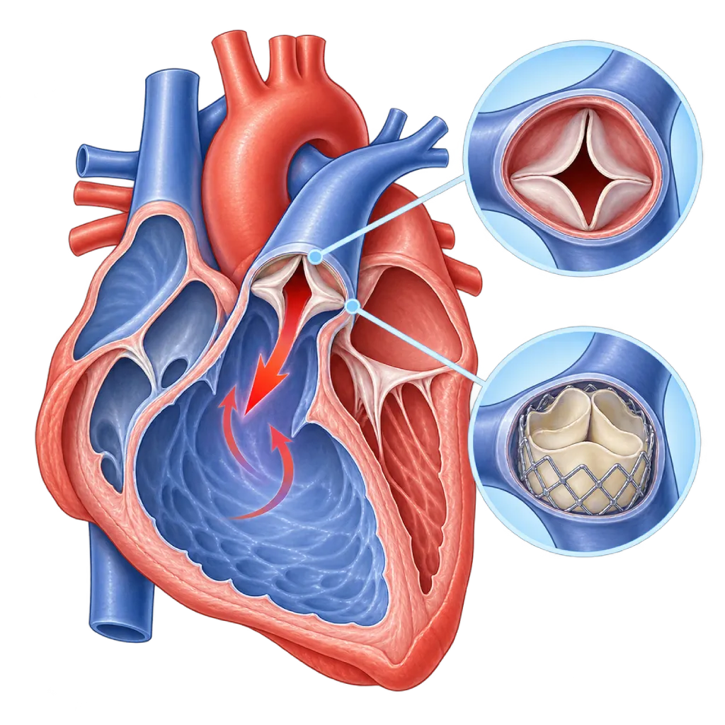
Pulmonary regurgitation is a leak in the pulmonary valve, so some of the blood pumped to the lungs flows back into the right ventricle. A mild leak is generally harmless, while a severe leak can strain the right ventricle.
SourcesSigns That Need Urgent Help
- Fainting or nearly fainting, especially during activity
- A very fast heartbeat with dizziness, chest pain, or shortness of breath
- Severe shortness of breath at rest
- Leg and abdominal swelling that increases quickly
- High fever and chills in people with an artificial pulmonary valve or conduit
If these signs appear, go to the nearest emergency department right away or call Heart Attack Center 1500 258.
01 / Understanding the condition
About Pulmonary Regurgitation
The pulmonary valve sits between the right ventricle and the pulmonary artery, the blood vessel that carries blood to the lungs to pick up oxygen. In pulmonary regurgitation, this valve does not close tightly, so some blood flows back into the right ventricle between heartbeats. Pressure on the right side of the heart is lower than on the left side, so this leak can often last a long time without symptoms.
A very mild leak is fairly often seen on echocardiography in healthy people and is not considered a disease. What needs attention is a moderate to severe leak. This condition belongs to the group of leaking heart valves, which is covered on the Valve Regurgitation / Insufficiency page.
Main Causes
A severe leak is usually found in people who have had congenital heart surgery, especially Tetralogy of Fallot repair. In this operation, the right ventricular outflow tract and the pulmonary valve are widened so that enough blood flows to the lungs, so the valve often no longer closes tightly. A leak can also develop after balloon widening of the valve for pulmonary stenosis. Other causes are high pressure in the lung blood vessels that widens the pulmonary artery, valve infection, and, rarely, a congenital valve abnormality or carcinoid syndrome.
Effects on the Right Ventricle
The right ventricle can cope with the leak for years by enlarging, so many people continue to feel well. However, if the enlargement continues, the right ventricle can weaken, the tricuspid valve can start leaking too, and heart rhythm disorders become more likely. A decline in exercise ability often develops slowly, so it goes unnoticed. That is why the size of the right ventricle and exercise capacity are monitored over time, rather than relying only on symptoms.
Monitoring and Treatment
The ESC 2020 and AHA/ACC 2018 guidelines for adult congenital heart disease recommend lifelong monitoring, including regular cardiac MRI to measure right ventricular volume and function. Pulmonary valve replacement is considered if symptoms appear, or without symptoms if the right ventricle enlarges beyond a certain limit, its function declines, or rhythm disorders appear. The valve can be replaced through surgery or through a catheter when the anatomy is suitable. If the leak is caused by pulmonary hypertension, treatment is directed at the underlying disease.
02 / Understanding the condition
Symptoms to Recognize.
Common Symptoms
Many people have no symptoms for years. When symptoms do appear, they usually develop slowly.
- Gradually declining exercise ability
- Shortness of breath during activity
- Tiring easily
- Palpitations or a heartbeat that feels irregular
- Swelling in the ankles or legs
- A bloated or swollen abdomen due to fluid buildup
- A heart murmur your doctor hears at a checkup
03 / Understanding the condition
What Causes This Condition?
- Previous Tetralogy of Fallot repair surgery that widened the right ventricular outflow tract
- Widening of the pulmonary valve with a balloon or surgery for pulmonary stenosis
- Pulmonary hypertension that widens the pulmonary artery and the valve ring
- Valve infection (infective endocarditis), including infection of an artificial valve or conduit
- Congenital abnormalities of the pulmonary valve, such as valve leaflets that are not fully formed
- Carcinoid syndrome, a rare condition that can damage the valves on the right side of the heart
- Declining function of an artificial pulmonary valve or conduit after many years
04 / Understanding the condition
Who Is at Higher Risk?
Medical
A history of surgery or catheter procedures for congenital heart disease, pulmonary hypertension, endocarditis, and having an artificial valve or conduit to the pulmonary artery.
Age
In people with Tetralogy of Fallot who had surgery as children, the effects of the leak on the right ventricle usually start to show in adolescence or young adulthood.
Genetic
The underlying congenital heart disease, such as Tetralogy of Fallot, can be linked to certain genetic abnormalities, such as chromosome 22q11 deletion.
Other
Injecting drugs and poor dental hygiene increase the risk of valve infection on the right side of the heart.
Groups at Higher Risk
- People with Repaired Tetralogy of Fallot: A leaking pulmonary valve after repair is common and needs lifelong monitoring. Covered on the Tetralogy of Fallot page.
- People with a Previously Widened Pulmonary Valve: Widening for pulmonary stenosis with a balloon or surgery can leave a leak that increases over time.
- People with Pulmonary Hypertension: High lung pressure widens the pulmonary artery so the valve does not close tightly.
- People with an Artificial Pulmonary Valve or Conduit: An artificial valve can wear out, narrow, leak, or become infected, so regular checkups are needed.
05 / Understanding the condition
How Is It Diagnosed?
The murmur of pulmonary regurgitation is often soft and easy to miss, so testing is usually guided by a history of congenital heart surgery or pulmonary hypertension. Echocardiography assesses the degree of leakage, the size and function of the right ventricle, tricuspid valve leakage, and estimated lung pressure.
Cardiac MRI is the reference test for pulmonary regurgitation because it can precisely measure right ventricular volume and the percentage of blood leaking back. These regular MRI results are used to determine the timing of valve replacement. An ECG and a rhythm recording over several days look for rhythm disorders, while cardiopulmonary exercise testing measures exercise ability objectively. A CT scan or cardiac catheterization is used when planning catheter-based valve replacement, including checking the position of the coronary arteries.
Echocardiography
Assesses the degree of leakage, the size and function of the right ventricle, tricuspid valve leakage, and estimated lung pressure.
Cardiac MRI
The reference test to measure right ventricular volume and function and the percentage of blood leaking back, the basis for timing valve replacement.
Electrocardiogram (ECG)
Looks for rhythm disorders and measures the width of the ventricles' electrical signal (QRS duration), which is monitored in people with Tetralogy of Fallot.
Holter Monitoring
Records the heart rhythm for 24 to 48 hours to look for rhythm disorders not caught on a standard ECG.
Cardiopulmonary exercise testing objectively measures exercise ability and oxygen use to track changes over time.
Cardiac CT Scan
Maps the right ventricular outflow tract and the position of the coronary arteries when planning catheter-based valve replacement.
06 / Understanding the condition
Treatment Options.
Treatment is decided by a doctor after an assessment. The categories below can help you follow the conversation with your doctor.
Regular Monitoring
Mild to moderate leaks are monitored with echocardiography, and in people with congenital heart disease also with regular cardiac MRI.
Surgical Pulmonary Valve Replacement
The pulmonary valve is replaced with a biological valve or a valved conduit through surgery, sometimes together with other repairs in the heart.
Transcatheter Pulmonary Valve Replacement
An artificial valve is placed through a catheter from a blood vessel in the groin or neck when the anatomy is suitable, without opening the chest.
Treatment of Rhythm Disorders
Medicines, ablation, or devices such as an ICD are considered based on the type of rhythm disorder and the doctor's risk assessment.
Treatment of the Underlying Disease
If the leak is caused by pulmonary hypertension, treatment is directed at the pulmonary hypertension itself.
Medicines for Symptoms
Diuretics can relieve swelling once fluid has built up. Your doctor determines the type and dose.
07 / Understanding the condition
Complications to Watch For.
Right Ventricular Enlargement and Reduced Function
Years of leakage cause the right ventricle to enlarge, and it can weaken if valve replacement is delayed.
Tricuspid Regurgitation
The enlarged right ventricle stretches the tricuspid valve ring, so this valve starts leaking too. Covered on the Tricuspid Regurgitation page.
Heart Rhythm Disorders
Rhythm disorders of the atria and of the ventricles occur more easily when the right ventricle is enlarged. Covered on the Heart Rhythm Disorders / Arrhythmia page.
Right Heart Failure
If the right ventricle weakens, leg swelling, abdominal bloating, and tiring quickly appear. Covered on the Heart Failure page.
Infective Endocarditis
An artificial valve or conduit in the pulmonary artery can become infected. Covered on the Infective Endocarditis page.
08 / Understanding the condition
What Can You Do?
- Keep up lifelong heart checkups if you have had congenital heart surgery
- Have regular cardiac MRI as your doctor recommends, even if you have no symptoms
- Keep your teeth and mouth clean and have regular dental checkups
- Ask your doctor whether you need preventive antibiotics before dental procedures if you have an artificial valve
- Stay physically active on a regular basis within the limits set by your doctor
- Report right away if your exercise ability declines or you notice palpitations
09 / Understanding the condition
When Should You See a Doctor?
If you have congenital heart disease and have had surgery, see a cardiologist regularly from childhood into adulthood, even if you feel well. Book a checkup earlier than scheduled if new symptoms appear. Seek help right away if you faint or have a very fast heartbeat with dizziness.
- Exercise ability that has declined compared with before
- Shortness of breath or tiring quickly during activity
- Palpitations or a heartbeat that feels irregular
- Leg swelling or a bloated abdomen
- Fever without a clear cause if you have an artificial pulmonary valve or conduit
- Planning a pregnancy if you are a woman with a history of congenital heart surgery
Don't Wait in an Emergency.
If symptoms are severe or come on suddenly, go to the nearest emergency department right away or call Heart Attack Center 1500 258, rather than waiting for an appointment.
10 / Common questions
Questions You May Have.
Frequently asked questions about pulmonary regurgitation.
Does mild pulmonary regurgitation need treatment?
A very mild pulmonary valve leak is often found in healthy people and does not need treatment or activity restrictions. A mild leak due to other causes only needs monitoring as your doctor advises. More attention is needed if the leak is moderate to severe, especially after congenital heart surgery or if pulmonary hypertension is present.
Why does a leak appear after Tetralogy of Fallot surgery?
In Tetralogy of Fallot repair surgery, the narrow right ventricular outflow tract is widened so that enough blood flows to the lungs. This widening often means the pulmonary valve no longer closes tightly. During childhood, the body usually tolerates the leak well, but its effect on the right ventricle needs to be monitored into adulthood. A full explanation is covered on the Tetralogy of Fallot page.
When does the pulmonary valve need to be replaced?
Replacement is considered if a severe leak causes symptoms, or without symptoms if cardiac MRI shows the right ventricle has enlarged beyond a certain limit, its function has declined, tricuspid leakage has increased, or rhythm disorders have appeared. The goal is to act before the right ventricle becomes hard to recover. The decision is made by the adult congenital heart team together with you.
What is the difference between catheter-based and surgical valve replacement?
With a catheter, the artificial valve is brought in from a blood vessel in the groin or neck and placed inside the old conduit or valve, without opening the chest, so recovery is usually shorter. This approach requires a conduit of suitable size and shape. Surgery is chosen if the anatomy is not suitable or if other repairs need to be done at the same time.
Can I exercise with pulmonary regurgitation?
In general, yes, and regular physical activity is recommended for people with congenital heart disease. Exercise limits are based on the size and function of the right ventricle, whether rhythm disorders are present, and the results of exercise testing. If you feel dizzy, have palpitations, or faint during exercise, stop the activity and get checked right away.
Can women with pulmonary regurgitation become pregnant?
Many women with pulmonary regurgitation can go through pregnancy well. A severe leak with a weakened right ventricle or rhythm disorders increases the risk during pregnancy, so an assessment before pregnancy is recommended. Sometimes valve replacement is considered first. Plan your pregnancy together with your cardiologist and your obstetrician.
Sources and Medical Review
Translated from the Indonesian edition medically reviewed by dr. Sri Diniharini, Sp.JP(K).
Sources as cited in the Indonesian edition.
- Baumgartner H, De Backer J, Babu-Narayan SV, Budts W, Chessa M, Diller GP, et al. 2020 ESC Guidelines for the management of adult congenital heart disease. Eur Heart J. 2021;42(6):563-645.
- Stout KK, Daniels CJ, Aboulhosn JA, Bozkurt B, Broberg CS, Colman JM, et al. 2018 AHA/ACC Guideline for the Management of Adults With Congenital Heart Disease: A Report of the American College of Cardiology/American Heart Association Task Force on Clinical Practice Guidelines. Circulation. 2019;139(14):e698-e800.
- Zoghbi WA, Adams D, Bonow RO, Enriquez-Sarano M, Foster E, Grayburn PA, et al. Recommendations for Noninvasive Evaluation of Native Valvular Regurgitation: A Report from the American Society of Echocardiography Developed in Collaboration with the Society for Cardiovascular Magnetic Resonance. J Am Soc Echocardiogr. 2017;30(4):303-371.
- Geva T. Repaired tetralogy of Fallot: the roles of cardiovascular magnetic resonance in evaluating pathophysiology and for pulmonary valve replacement decision support. J Cardiovasc Magn Reson. 2011;13(1):9.
- van der Ven JPG, van den Bosch E, Bogers AJCC, Helbing WA. Current outcomes and treatment of tetralogy of Fallot. F1000Res. 2019;8:F1000 Faculty Rev-1530.
- Cheatham JP, Hellenbrand WE, Zahn EM, Jones TK, Berman DP, Vincent JA, et al. Clinical and Hemodynamic Outcomes up to 7 Years After Transcatheter Pulmonary Valve Replacement in the US Melody Valve Investigational Device Exemption Trial. Circulation. 2015;131(22):1960-1970.
- McElhinney DB, Zhang Y, Levi DS, Georgiev S, Biernacka EK, Goldstein BH, et al. Reintervention and Survival After Transcatheter Pulmonary Valve Replacement. J Am Coll Cardiol. 2022;79(1):18-32.
Doctors Who Treat This Condition

dr. Radityo Prakoso, Sp.JP(K)
Pediatrics & Congenital Heart Disease
Congenital and Structural Heart Center
Clinical focusPediatric Cardiology Consultation · Congenital Heart Disease Care
View Profile
dr. Salomo Purba, Sp.BTKV, Subsp.JPK(K)
Pediatric and Congenital Heart Surgery
Congenital and Structural Heart Center +2 centers
Clinical focusPediatric Heart Surgery · Congenital Heart Defect Surgery
View Profile
dr. Sri Diniharini, Sp.JP(K)
Echocardiography & Cardiovascular Imaging
Cardiovascular Diagnostic Center +1 center
Clinical focusAdvanced Echocardiography · Transesophageal Echocardiography
View Profile