
Quick answer
What Is Transposition of the Great Arteries?
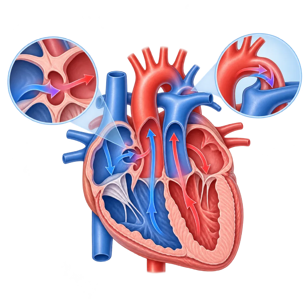
Transposition of the great arteries is a congenital heart defect in which the aorta and the pulmonary artery have switched places. Oxygen-rich blood circulates back to the lungs, so the baby looks blue from the first day and usually needs surgery within the first few weeks.
Sources01 / Understanding the condition
About Transposition of the Great Arteries
In a normal heart, the right ventricle pumps oxygen-poor blood to the lungs through the pulmonary artery, then the left ventricle pumps oxygen-rich blood to the body through the aorta. In transposition of the great arteries (often shortened to TGA), the positions of the two great arteries are switched: the aorta comes out of the right ventricle and the pulmonary artery comes out of the left ventricle. As a result, two separate circuits form. Blood from the body returns to the body without picking up oxygen, while blood from the lungs returns to the lungs. This condition is found in about 5 of every 100 congenital heart defects and is more common in baby boys. The defect forms in the early weeks of pregnancy and is not caused by anything the parents did.
Why a Baby Can Survive for a While
A baby can only survive if blood from the two circuits mixes through shortcuts: the foramen ovale or a hole in the wall between the atria, the ductus arteriosus, or a hole in the wall between the ventricles if there is one. The foramen ovale and the ductus tend to close within a few days after birth, so the baby can become very blue within hours. Prostaglandin medicines and a catheter procedure to widen the hole in the wall between the atria (balloon atrial septostomy) keep the blood mixing until surgery.
Arterial Switch Operation
The main treatment is the arterial switch operation, usually within the first two to three weeks of life. The aorta and the pulmonary artery are moved to the correct ventricles, and the coronary arteries are also moved to the new aorta. Long-term results are good, and the majority of children grow up with normal activity. Adults who had surgery before the arterial switch era usually had an atrial switch operation (Mustard or Senning). They need special monitoring because their right ventricle works as the main pump for the body.
Another Form: Congenitally Corrected Transposition
There is another, rarer form in which the ventricles are also switched, so blood still flows in the right sequence and the baby is not blue. This form is often only found in adulthood because of rhythm disorders, a slow heart rate, or valve leaking. The ESC 2020 and AHA/ACC 2018 guidelines recommend lifelong monitoring for both forms. An overview of other congenital heart defects is covered on the Congenital Heart Disease page.
02 / Understanding the condition
Symptoms to Recognize.
Common Symptoms
- Skin, lips, and tongue look blue from a few hours after birth
- Blueness that does not improve even with oxygen
- Fast breathing even though the baby does not seem severely short of breath
- Difficulty feeding or tiring quickly while feeding
- Low oxygen saturation on newborn screening
Symptoms in Newborns
If there is a large hole in the wall between the ventricles, the blueness may be mild and signs of heart failure may only appear within a few weeks.
- Blueness that gets worse during the first one to three days
- The baby looks sluggish or less active
- Fast breathing and sweating while feeding
- Difficulty gaining weight in the first few weeks
Symptoms in Children
After an arterial switch operation, the majority of children have no complaints at all; the following symptoms should be checked.
- Chest pain or paleness during strenuous play
- Fainting during activity
- Tiring more quickly than peers
- A new heart murmur at a checkup
Symptoms in Adults
Especially in adults who had an atrial switch operation in the past or who have congenitally corrected transposition.
- Tiring quickly and shortness of breath during activity
- Palpitations or an irregular rhythm
- Swelling in the legs or abdomen
- Dizziness, a heartbeat that feels slow, or fainting
03 / Understanding the condition
What Causes This Condition?
- A problem with the rotation and division of the outflow tracts of the fetal heart in the early weeks of pregnancy
- The exact cause is usually unknown and is rarely linked to a genetic syndrome
- Uncontrolled diabetes in the mother before pregnancy is linked to a higher risk
- Changes in genes that control the left-right orientation of the body in a small number of cases
04 / Understanding the condition
Who Is at Higher Risk?
Medical
Uncontrolled diabetes in the mother that was present before pregnancy, as well as some medicines and infections in the first trimester, are linked to a higher risk of congenital heart defects.
Genetic
A history of congenital heart disease in a parent or sibling slightly increases the risk. Unlike other defects, transposition is rarely accompanied by a chromosomal syndrome.
Other
Transposition is found more often in baby boys, about twice as often as in baby girls.
Lifestyle
Smoking and drinking alcohol during pregnancy increase the risk of congenital heart defects in general.
Groups at Higher Risk
- Baby Boys: Transposition is found about twice as often in baby boys as in baby girls.
- Babies of Mothers with Diabetes: Uncontrolled diabetes that was present before pregnancy is linked to a risk of transposition and other congenital heart defects.
- Children and Adults After Arterial Switch: The aortic root, aortic valve, pulmonary artery, and coronary arteries need monitoring because problems can develop years later.
- Adults After Mustard or Senning Surgery: The right ventricle that pumps to the body can weaken, and atrial arrhythmias and sinus node problems often appear.
05 / Understanding the condition
How Is It Diagnosed?
Transposition may be suspected on a pregnancy ultrasound if the two great arteries appear to leave the fetal heart side by side, and it is then confirmed with fetal echocardiography. Diagnosis before birth is linked to a better condition of the baby at the time of surgery, because delivery and medicines can be prepared in advance.
After birth, the main clues are blueness that does not improve with oxygen and low oxygen saturation on newborn screening. Echocardiography confirms the position of the great arteries and assesses the size of the holes in the walls between the atria and between the ventricles, the ductus arteriosus, the valves, and the pattern of the coronary arteries, which is important for surgical planning. A chest X-ray and an ECG complete the assessment. Cardiac catheterization is done when a balloon septostomy or additional information is needed.
After surgery, monitoring uses echocardiography, cardiac MRI or CT, and an ECG. If there is chest pain or fainting during activity, the doctor also assesses coronary blood flow.
Fetal echocardiography: confirms suspected transposition from a pregnancy ultrasound so the place of delivery and medicines can be prepared in advance.
Pulse oximetry screening: measures oxygen saturation in the right hand and a foot of a newborn before going home from the birthing facility.
Echocardiography
Confirms the position of the great arteries, the size of the holes where blood mixes, valve function, and the coronary artery pattern for surgical planning.
Cardiac Catheterization
Used for balloon atrial septostomy, which widens the hole in the wall between the atria so more blood can mix.
Cardiac MRI
Assesses the aortic root, pulmonary artery, and ventricular function during long-term monitoring after surgery.
Electrocardiogram (ECG)
Looks for signs of reduced coronary blood flow after an arterial switch and rhythm disorders after an atrial switch.
Cardiac CT Scan
Maps the coronary arteries and pulmonary artery when there are symptoms or before a repeat procedure.
06 / Understanding the condition
Treatment Options.
Treatment is decided by a doctor after an assessment. The categories below can help you follow the conversation with your doctor.
Prostaglandin and Intensive Care
Prostaglandin medicine given by infusion keeps the ductus open so blood can mix, with close monitoring in the neonatal intensive care unit.
Balloon Atrial Septostomy
A balloon at the tip of a catheter is pulled across the wall between the atria to widen the hole, so more oxygen-rich blood reaches the body.
Arterial Switch Operation
The main operation in the early weeks: the aorta and the pulmonary artery are moved to the correct ventricles along with the coronary arteries. Covered on the Congenital Heart Surgery page.
Other Operations Based on Anatomy
If there is a hole in the wall between the ventricles and narrowing of the path to the lungs, the doctor may choose a Rastelli operation or a similar technique that uses a tube to the pulmonary artery.
Arrhythmia and Heart Failure Treatment
In adults after an atrial switch, doctors may use medicines, ablation, a pacemaker, or an ICD as needed, along with monitoring of right ventricular function.
Lifelong Follow-Up
Regular echocardiography, MRI or CT, ECG, and exercise testing at an adult congenital heart disease service, as recommended by ESC 2020 and AHA/ACC 2018.
07 / Understanding the condition
Complications to Watch For.
Severe Lack of Oxygen in Babies
If blood mixing is not enough, oxygen levels drop sharply and acid builds up in the body, so organs can be damaged within a short time.
Pulmonary Artery Narrowing
After an arterial switch, the pulmonary artery can narrow at the connection site and sometimes needs to be widened with a balloon or surgery.
Aortic Root Enlargement and Valve Leaking
The new aortic root can enlarge and its valve can leak over time, so its size is monitored regularly.
Coronary Artery Problems
The moved coronary arteries can narrow or kink. Chest pain or fainting during activity needs to be checked right away.
Systemic Right Ventricular Failure
After an atrial switch or in congenitally corrected transposition, the right ventricle that pumps to the body can weaken. Covered on the Heart Failure page.
Rhythm and Sinus Node Disorders
Atrial flutter, a slow heart rate, or heart block often appear after an atrial switch and may need medicines, ablation, or a pacemaker.
08 / Understanding the condition
What Can You Do?
- Transposition cannot be reliably prevented, but a healthy pregnancy helps lower the risk of congenital heart defects
- Control diabetes before and during pregnancy, take folic acid as recommended, and avoid smoking and alcohol
- Have scheduled pregnancy ultrasounds; diagnosis before birth helps prepare for delivery and the baby's care
- Make sure your baby has oxygen saturation screening before going home from the birthing facility
- Keep up regular checkups after surgery even if your child seems healthy, including during the move to adult doctors
- Report chest pain, fainting, or palpitations during activity in a child or adult after surgery
09 / Understanding the condition
When Should You See a Doctor?
A baby who looks blue, especially in the first days of life, needs to be taken to the emergency room right away. Blueness in a newborn is not something to wait out at home. Make an appointment with a cardiologist if:
- A pregnancy ultrasound shows a problem with the baby's great arteries.
- The baby's oxygen saturation screening result is abnormal.
- A baby with transposition and a hole in the wall between the ventricles breathes fast, sweats while feeding, or has trouble gaining weight.
- A child who has had surgery has chest pain, looks pale, or faints while playing.
- An adult with a history of atrial switch or corrected transposition often has palpitations, tires quickly, or has swelling.
- You have not had a checkup for a long time after surgery.
10 / Common questions
Questions You May Have.
Frequently asked questions about transposition of the great arteries.
Why does a baby with transposition need surgery so soon?
Without surgery, oxygen-rich blood keeps circulating through the lungs and not enough reaches the body. The natural shortcuts that help blood mix tend to close after birth. In addition, the left ventricle, which pumps to the low-pressure lungs, will weaken if surgery is delayed too long, so it is no longer ready to pump to the body. That is why the arterial switch operation is usually done within the first two to three weeks.
What is a balloon atrial septostomy?
A balloon atrial septostomy is a catheter procedure to widen the hole in the wall between the two atria. A balloon-tipped catheter is inserted through a blood vessel in the belly button or groin, then the balloon is inflated and pulled across the wall. The wider hole lets more oxygen-rich blood mix and reach the body. This procedure serves as a bridge until the baby is ready for the main operation.
What is life like for a child after an arterial switch operation?
The majority of children grow up, go to school, and play sports like their peers. Even so, the aortic root and its valve, the pulmonary artery, and the coronary arteries need regular monitoring because problems can develop later in life. Some children also need developmental support, for example with speech or learning. Routine checkups, and checks whenever there is chest pain or fainting during activity, help find problems early.
What is the difference between an arterial switch and an atrial switch operation?
An arterial switch operation moves the aorta and the pulmonary artery to the correct ventricles, so the left ventricle once again pumps to the body. An atrial switch operation, which was widely done before the 1990s, redirects blood flow at the level of the atria, so the right ventricle still pumps to the body. Adults with a history of atrial switch more often have arrhythmias and reduced right ventricular function, so their monitoring is closer.
Can women with a history of transposition get pregnant?
Many women with a history of transposition can get pregnant, but they need to be assessed first by a cardiologist who understands congenital heart disease. The assessment covers the function of the ventricle that pumps to the body, valve leaking, heart rhythm, and the medicines being taken. After an atrial switch, pregnancy can increase the load on the right ventricle and trigger arrhythmias. Plan your pregnancy with a cardiologist and an obstetrician.
Sources and Medical Review
Translated from the Indonesian edition medically reviewed by dr. Salomo Purba, Sp.BTKV, Subsp.JPK(K).
Sources as cited in the Indonesian edition.
- Baumgartner H, De Backer J, Babu-Narayan SV, et al. 2020 ESC Guidelines for the management of adult congenital heart disease. Eur Heart J. 2021;42(6):563-645.
- Stout KK, Daniels CJ, Aboulhosn JA, et al. 2018 AHA/ACC Guideline for the Management of Adults With Congenital Heart Disease: A Report of the American College of Cardiology/American Heart Association Task Force on Clinical Practice Guidelines. Circulation. 2019;139(14):e698-e800.
- Villafañe J, Lantin-Hermoso MR, Bhatt AB, et al. D-transposition of the great arteries: the current era of the arterial switch operation. J Am Coll Cardiol. 2014;64(5):498-511.
- Warnes CA. Transposition of the great arteries. Circulation. 2006;114(24):2699-2709.
- Khairy P, Clair M, Fernandes SM, et al. Cardiovascular outcomes after the arterial switch operation for D-transposition of the great arteries. Circulation. 2013;127(3):331-339.
- Bonnet D, Coltri A, Butera G, et al. Detection of transposition of the great arteries in fetuses reduces neonatal morbidity and mortality. Circulation. 1999;99(7):916-918.
- Martin GR, Ewer AK, Gaviglio A, et al. Updated Strategies for Pulse Oximetry Screening for Critical Congenital Heart Disease. Pediatrics. 2020;146(1):e20191650.
Doctors Who Treat This Condition

dr. Radityo Prakoso, Sp.JP(K)
Pediatrics & Congenital Heart Disease
Congenital and Structural Heart Center
Clinical focusPediatric Cardiology Consultation · Congenital Heart Disease Care
View Profile
dr. Salomo Purba, Sp.BTKV, Subsp.JPK(K)
Pediatric and Congenital Heart Surgery
Congenital and Structural Heart Center +2 centers
Clinical focusPediatric Heart Surgery · Congenital Heart Defect Surgery
View Profile
dr. Tartila, Sp.A, Subsp.ETIA(K)
Pediatric Emergency and Intensive Care
Congenital and Structural Heart Center
Clinical focusPediatric Intensive Care · Pediatric Emergency Care
View Profile