
Quick answer
What Is Total Anomalous Pulmonary Venous Return?
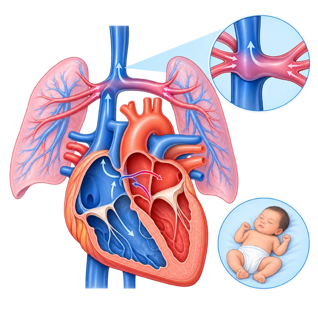
Total anomalous pulmonary venous return is a congenital defect in which all four pulmonary veins drain not into the left atrium but into the right side of the heart. Babies look blue or have trouble breathing and need surgery in infancy.
Sources01 / Understanding the condition
About Total Anomalous Pulmonary Venous Return
Normally, four lung veins (pulmonary veins) carry oxygen-rich blood from the lungs to the left atrium, and the blood is then pumped to the rest of the body. In total anomalous pulmonary venous return (TAPVR), none of the four pulmonary veins is connected to the left atrium. Instead, these veins join together and drain into the right atrium or into a large vein that flows to the right atrium. As a result, oxygen-rich blood from the lungs mixes with oxygen-poor blood from the body on the right side of the heart.
To reach the body, this mixed blood has to cross into the left atrium through an opening in the wall between the atria, namely the foramen ovale or an atrial septal defect. This opening becomes a lifeline. Because the blood circulating to the body is mixed, the baby's oxygen level is lower than normal and the skin looks bluish.
Four Types by Drainage Site
In the supracardiac type, the pulmonary veins drain into a large vein above the heart. In the cardiac type, the pulmonary veins drain directly into the right atrium or into the coronary sinus. In the infracardiac type, the flow goes down through the diaphragm to a vein in the abdomen or liver. The mixed type combines several drainage sites. The infracardiac type is more often accompanied by obstruction.
Obstructed and Unobstructed Forms
The impact of this defect depends largely on whether the pulmonary venous pathway is obstructed. If it is obstructed, blood backs up in the lungs, making them wet and stiff. The baby looks very blue and short of breath within the first hours to days and needs surgery right away. If it is not obstructed, the baby may look only slightly blue, breathe fast, have difficulty feeding, and gain weight poorly over the first weeks to months. Both forms still need surgery.
After Surgery
Surgery connects the confluence of pulmonary veins to the left atrium, closes the abnormal pathway, and closes the opening in the atrial wall. During surgery, a heart-lung machine temporarily takes over the work of the heart and lungs. Many children grow and stay active well after surgery. Some children develop narrowing of the pulmonary veins after surgery, usually within the first few months, so regular follow-up is still needed into adulthood.
02 / Understanding the condition
Symptoms to Recognize.
Common Symptoms
- Bluish skin, lips, or nails
- Fast or labored breathing
- Difficulty feeding and tiring quickly while feeding
- Poor weight gain
- Low oxygen level when measured with an oximeter
Symptoms in Newborns
Especially in the obstructed form, symptoms appear within the first hours to days after birth.
- Obvious blueness within a few hours after birth
- Very fast breathing with chest wall retractions
- Grunting when breathing
- Blueness that does not improve with regular oxygen
Symptoms in Infants
In the unobstructed form, symptoms often appear gradually over the first weeks to months.
- Fast breathing and sweating while feeding
- Poor weight gain
- Frequent coughs and colds or pneumonia
- Mild blueness that is easily missed
03 / Understanding the condition
What Causes This Condition?
- The pulmonary veins fail to connect to the left atrium while the fetal heart and lungs are forming
- Temporary connections between the pulmonary veins and the body's veins during fetal life persist
- In the majority of babies, the exact trigger is unknown
- Genetic factors in some families, including heterotaxy syndrome (an unusual arrangement of the organs)
- It is sometimes accompanied by other congenital heart defects
04 / Understanding the condition
Who Is at Higher Risk?
Genetic
Heterotaxy syndrome, an unusual arrangement of the chest and abdominal organs, is often accompanied by abnormal pulmonary venous drainage. A family history of congenital heart disease (CHD) also increases the likelihood.
Medical
Maternal factors linked to CHD in general, such as uncontrolled diabetes or infection in early pregnancy, may play a role, although the evidence for this particular defect is limited.
Other
This defect is hard to see on a pregnancy ultrasound because blood flow to the fetal lungs is still low, so many cases are only found after birth.
Groups at Higher Risk
- Babies with the Obstructed Form: They need surgery right after diagnosis because their condition can worsen within hours.
- Babies with Heterotaxy Syndrome: They often have several heart defects at once, so their treatment is more complex.
- Children Who Have Had Surgery: They need regular follow-up to detect pulmonary vein narrowing, rhythm disorders, and high lung pressure.
05 / Understanding the condition
How Is It Diagnosed?
This defect may be suspected when newborn pulse oximetry screening shows a low oxygen level, or when the baby looks blue and short of breath. The obstructed form often resembles severe lung disease in newborns, so doctors need to be alert when the oxygen level does not improve despite breathing support.
Echocardiography confirms the diagnosis by showing that the pulmonary veins do not drain into the left atrium, where they drain instead, and whether there is an obstruction. This test also assesses the size of the opening in the atrial wall and the lung pressure. Cardiac CT angiography helps map the path of the pulmonary veins before surgery. This defect is hard to see on a pregnancy ultrasound, so screening after birth is still important even if the pregnancy ultrasound was normal.
Pulse oximetry screening after 24 hours of age finds low oxygen levels before the baby looks seriously ill.
Echocardiography
The main test to see where all four pulmonary veins drain, whether there is an obstruction, the size of the opening in the atrial wall, and the lung pressure.
Chest X-Ray
In the obstructed form, the lungs look wet while the heart is not enlarged, so it is easily mistaken for lung disease.
Cardiac CT Scan
Maps the path and drainage of the pulmonary veins in three dimensions before surgery when echocardiography is not clear enough.
Electrocardiogram (ECG)
Assesses the strain on the right side of the heart and the heart rhythm.
Cardiac Catheterization
Rarely needed for diagnosis. Used when the anatomy is unclear or to treat pulmonary vein narrowing after surgery.
06 / Understanding the condition
Treatment Options.
Treatment is decided by a doctor after an assessment. The categories below can help you follow the conversation with your doctor.
Stabilization in Intensive Care
The baby is given breathing support, medicines to support the heart, and correction of the blood acid balance while being prepared for surgery. This stabilizes the baby but does not correct the defect.
Corrective Surgery
The confluence of pulmonary veins is connected directly to the left atrium, the abnormal pathway is tied off, and the opening in the atrial wall is closed, with the help of a heart-lung machine. Covered on the Congenital Heart Surgery page.
Urgent Surgery for the Obstructed Form
The obstructed form is an infant heart surgery emergency and is operated on right after the diagnosis is confirmed.
Treatment of Pulmonary Vein Narrowing
Narrowing after surgery can be treated with another operation or with widening through a catheter, depending on its location and severity.
Long-Term Follow-Up
Regular echocardiography and ECGs assess the pulmonary veins, lung pressure, and heart rhythm into adulthood.
07 / Understanding the condition
Complications to Watch For.
Respiratory Failure in Newborns
In the obstructed form, blood backs up in the lungs, so the baby becomes severely short of breath and needs breathing support and surgery right away.
Pulmonary Hypertension
Obstruction and excess flow raise the pressure in the lung vessels, both before and after surgery. Covered on the Pulmonary Hypertension page.
Pulmonary Vein Narrowing After Surgery
Some children develop narrowing at the connection or inside the pulmonary veins, usually within the first few months, and may need another procedure.
Heart Rhythm Disorders
Rhythm disorders arising from the atria can appear after surgery, so ECG and rhythm monitoring are part of follow-up.
Right Heart Failure
The right side of the heart receives all the returning blood, so it has to work hard. Without surgery, the baby is at risk of heart failure and failure to thrive.
08 / Understanding the condition
What Can You Do?
- Have regular prenatal checkups, including an ultrasound that assesses the baby's heart
- Make sure your newborn has pulse oximetry screening before going home
- Watch your baby's skin color, breathing, and feeding in the first few weeks
- Control your blood sugar before and during pregnancy if you have diabetes
- After surgery, keep to the follow-up schedule even if your child seems healthy
09 / Understanding the condition
When Should You See a Doctor?
This defect needs prompt treatment. Take your baby to the emergency room as quickly as possible if you notice the following signs, especially in the first days and weeks after birth. For a child who has already had surgery, contact the doctor before the scheduled follow-up if symptoms come back.
- Your baby's lips, tongue, or skin look blue or grayish
- Very fast breathing, grunting, or the chest pulling inward
- Your baby refuses to feed or is very limp
- Your baby's oximetry result is low or the baby did not pass the screening
- A child who has had surgery is breathing fast again, coughing often, or has stopped gaining weight
10 / Common questions
Questions You May Have.
Questions parents often ask about total anomalous pulmonary venous return.
Can this defect be seen on a pregnancy ultrasound?
Sometimes, but it is often missed. Before birth, blood flow to the fetal lungs is still low, so the abnormal drainage of the pulmonary veins is hard to see, even with fetal echocardiography. That is why many babies are only diagnosed after birth. Pulse oximetry screening in newborns and watching for blueness or fast breathing help find this defect earlier.
When does a baby with this defect need surgery?
Babies with the obstructed form have surgery right after the diagnosis is confirmed, because their condition can worsen within hours. Babies with the unobstructed form usually have surgery in the first weeks or months, once their condition is stable, because waiting too long adds strain on the right side of the heart and the lungs. The medical team will explain the right timing for your baby.
How do children do after surgery?
Many children grow and stay active well after corrective surgery. The problem to watch for is narrowing of the pulmonary veins, which usually appears within the first few months after surgery. That is why echocardiography is done fairly often in the first year, and then regularly into adulthood. Rhythm disorders and high lung pressure are also monitored during follow-up.
What warning signs should I watch for after my child comes home from surgery?
Watch for breathing that becomes fast or labored again, a cough that does not go away, blueness, tiring quickly while feeding or playing, and weight that stops increasing. These signs can point to pulmonary vein narrowing or high lung pressure. Contact the doctor right away, or take your child to the emergency room if the breathing difficulty is severe. Do not wait for the next scheduled follow-up if symptoms appear.
Are younger siblings of a child with this defect at the same risk?
The risk of a heart defect in siblings is slightly higher than in families without a history of congenital heart disease, but the majority of younger siblings are born with a normal heart. In a later pregnancy, the obstetrician may recommend fetal echocardiography. If this defect occurs with heterotaxy syndrome or other abnormalities, genetic counseling can help the family understand the risk.
Sources and Medical Review
Translated from the Indonesian edition medically reviewed by dr. Radityo Prakoso, Sp.JP(K).
Sources as cited in the Indonesian edition.
- Seale AN, Uemura H, Webber SA, et al. Total anomalous pulmonary venous connection: morphology and outcome from an international population-based study. Circulation. 2010;122(25):2718-26.
- Shi G, Zhu Z, Chen J, et al. Total anomalous pulmonary venous connection: the current management strategies in a pediatric cohort of 768 patients. Circulation. 2017;135(1):48-58.
- Harada T, Nakano T, Oda S, et al. Surgical results of total anomalous pulmonary venous connection repair in 256 patients. Interact Cardiovasc Thorac Surg. 2019;28(3):421-6.
- Martin GR, Ewer AK, Gaviglio A, et al. Updated strategies for pulse oximetry screening for critical congenital heart disease. Pediatrics. 2020;146(1):e20191650.
- Donofrio MT, Moon-Grady AJ, Hornberger LK, et al. Diagnosis and treatment of fetal cardiac disease: a scientific statement from the American Heart Association. Circulation. 2014;129(21):2183-242.
- Pierpont ME, Brueckner M, Chung WK, et al. Genetic basis for congenital heart disease: revisited: a scientific statement from the American Heart Association. Circulation. 2018;138(21):e653-e711.
Doctors Who Treat This Condition

dr. Salomo Purba, Sp.BTKV, Subsp.JPK(K)
Pediatric and Congenital Heart Surgery
Congenital and Structural Heart Center +2 centers
Clinical focusPediatric Heart Surgery · Congenital Heart Defect Surgery
View Profile
dr. Tartila, Sp.A, Subsp.ETIA(K)
Pediatric Emergency and Intensive Care
Congenital and Structural Heart Center
Clinical focusPediatric Intensive Care · Pediatric Emergency Care
View Profile
dr. Radityo Prakoso, Sp.JP(K)
Pediatrics & Congenital Heart Disease
Congenital and Structural Heart Center
Clinical focusPediatric Cardiology Consultation · Congenital Heart Disease Care
View Profile