
Quick answer
What Is Restrictive Cardiomyopathy?
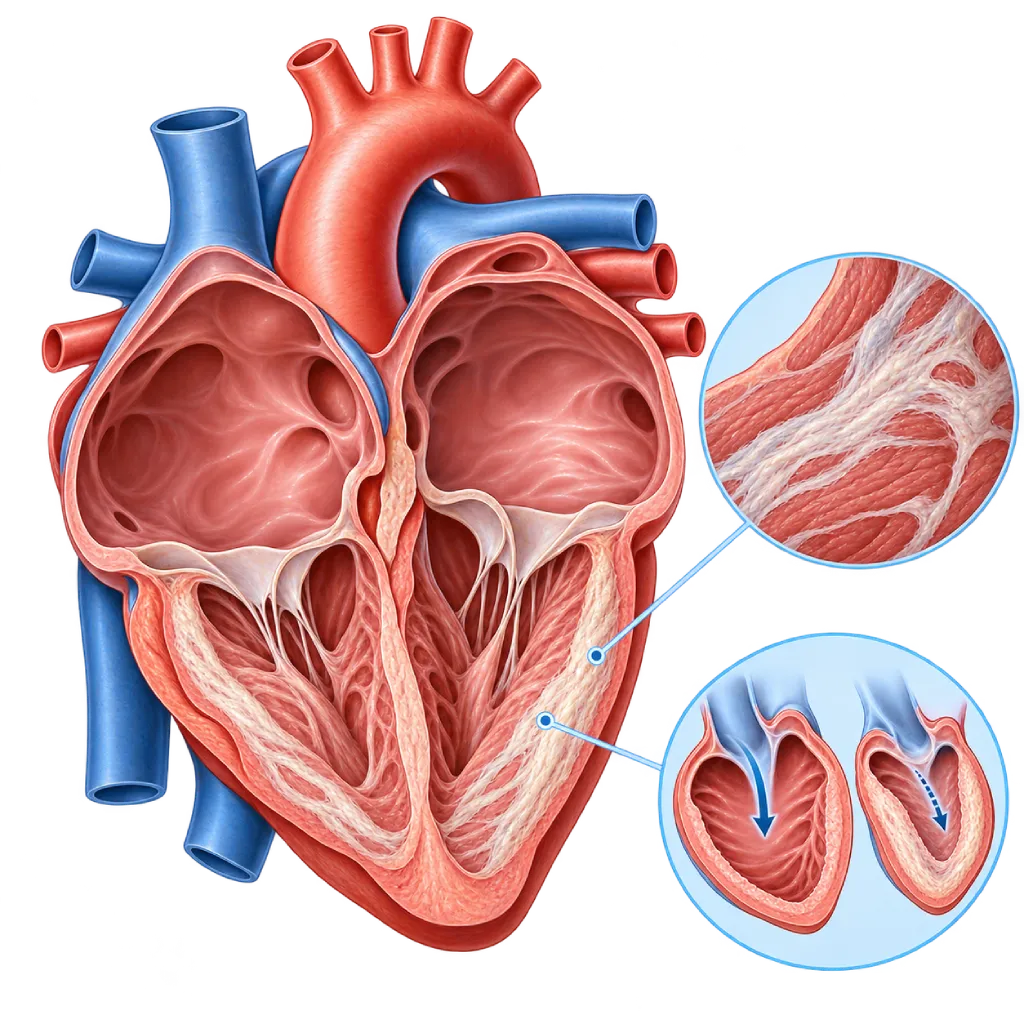
Restrictive cardiomyopathy is a heart muscle disease that makes the walls of the heart's chambers stiff, so the heart has trouble filling with blood even though its pumping strength is often still normal. This leads to shortness of breath, swelling, and heart failure.
SourcesSigns That Need Urgent Help
- Severe shortness of breath at rest or being unable to lie flat
- Sudden fainting
- A very fast heartbeat with severe dizziness
- Persistent chest pain
- Swelling that increases quickly in the legs, abdomen, or whole body
- Sudden facial drooping, arm weakness, or slurred speech
If these signs appear, go to the nearest emergency department right away or call Heart Attack Center 1500 258.
01 / Understanding the condition
About Restrictive Cardiomyopathy
A healthy heart expands flexibly to take in blood, then pumps it throughout the body. In restrictive cardiomyopathy, the muscle of the heart's lower chambers (ventricles) becomes stiff and cannot expand well. Pressure inside the heart rises, blood backs up into the lungs and the body, and both upper chambers of the heart (atria) become enlarged. Pumping strength is often still normal in the early stages, so this condition is one of the causes of heart failure with preserved ejection fraction.
Because the ventricles cannot hold enough blood, the amount of blood pumped with each beat becomes small, especially when the heart beats fast during activity. That is why shortness of breath and fatigue are often first felt during movement.
A Rare Type of Cardiomyopathy
Restrictive cardiomyopathy is a rare type of cardiomyopathy. The 2023 European Society of Cardiology (ESC) guidelines use this term for a stiff heart with normal or small ventricles and walls that are not thickened. Storage diseases such as amyloidosis can cause similar stiffness, but usually with thickened walls. This group of storage diseases is covered on the Infiltrative Cardiomyopathy page.
Varied Causes
Causes can include changes in the genes for heart muscle proteins, especially in children and young adults; buildup of protein, iron, or other substances in the heart muscle; scar tissue after chest radiation or certain cancer drugs; and diseases of the heart's inner lining, such as endomyocardial fibrosis and inflammation caused by high levels of eosinophils, a type of white blood cell. In some people no cause is found, and this is called idiopathic.
Similar to Constrictive Pericarditis
Its symptoms and test results are very similar to those of constrictive pericarditis, a stiffening and thickening of the sac that surrounds the heart. Telling the two apart is important because constrictive pericarditis can be treated with surgery, while restrictive cardiomyopathy cannot. That condition is covered on the Constrictive Pericarditis page.
Treatment Approach
Treatment focuses on controlling fluid with carefully adjusted water pills (diuretics), controlling heart rhythm, preventing blood clots, and treating the cause when there is one, such as amyloidosis or iron overload. In advanced stages, a heart transplant may be considered. An overview of this family of diseases is covered on the Cardiomyopathy page.
02 / Understanding the condition
Symptoms to Recognize.
Common Symptoms
- Shortness of breath during activity, and over time also at rest
- Tiring easily and a reduced ability to be active
- Swelling in the feet and ankles
- An enlarged or full-feeling abdomen due to fluid
- Visibly bulging veins in the neck
- A pounding or irregular heartbeat
- Dizziness or fainting during activity
Symptoms in Children
In children, symptoms are often mistaken for asthma or a lung infection, so diagnosis is frequently delayed.
- Tiring quickly and being unable to keep up with children of the same age during play
- Shortness of breath or wheezing that does not improve with asthma medicine
- Fainting or chest pain during activity
- Difficulty gaining weight or slowed growth
- A swollen abdomen due to an enlarged liver or fluid
03 / Understanding the condition
What Causes This Condition?
- Changes in the genes for heart muscle proteins, including those inherited in families
- Buildup of amyloid protein in the heart muscle (amyloidosis)
- Buildup of excess iron (hemochromatosis) or other substances in the heart muscle
- Granulomatous inflammation, such as sarcoidosis, that leaves scar tissue
- Scar tissue after chest radiation or certain cancer drugs
- Endomyocardial fibrosis and inflammation caused by high eosinophil levels
- Idiopathic, when no cause is found despite a complete workup
04 / Understanding the condition
Who Is at Higher Risk?
Genetic
A family history of cardiomyopathy, sudden death at a young age, or hereditary amyloidosis increases the likelihood of a genetic cause.
Medical
Blood disorders such as multiple myeloma, iron overload, sarcoidosis, and high eosinophil levels can affect the heart muscle and make it stiff.
Age
Causes differ by age: genetic forms are more common in children and young adults, while amyloidosis is more common in older adults.
Other
Radiation to the chest area and certain cancer drugs, such as anthracyclines, can cause scar tissue in the heart years later.
Groups at Higher Risk
- Family Members of People with Cardiomyopathy: Close relatives of patients with a genetic form are advised to have a heart checkup and genetic counseling even if they have no symptoms.
- Cancer Survivors with Chest Radiation: Radiation and certain cancer drugs can make the heart stiff years later, so long-term monitoring is recommended.
- People with Certain Blood Disorders: Multiple myeloma and plasma cell disorders can cause amyloidosis, while high eosinophil levels can damage the heart's inner lining.
- Older Adults with Heart Failure: Heart failure with a still-normal pumping function in older adults is sometimes caused by amyloidosis, which makes the heart stiff.
05 / Understanding the condition
How Is It Diagnosed?
Your doctor asks about your symptoms, your history of cancer treatment and blood disorders, and any heart disease in your family, then checks for signs of congestion such as bulging neck veins, an enlarged liver, and swelling. An ECG and an NT-proBNP test are the first steps.
Echocardiography shows enlarged atria, ventricles that are not enlarged, and a stiff filling pattern. Cardiac MRI assesses the heart muscle tissue and helps find the cause, such as amyloidosis, iron deposits, or scar tissue. To distinguish the condition from constrictive pericarditis, your doctor may perform cardiac catheterization to measure the pressures on both sides of the heart at the same time. If the cause is still unclear, a heart muscle biopsy, a bone scan for transthyretin amyloidosis, blood and urine protein tests, and genetic testing may be considered. The results of all these tests determine whether there is a specific treatment for the cause.
Echocardiography
Assesses the size of the atria and ventricles, wall thickness, the stiff filling pattern, and lung pressure, and helps distinguish the condition from constrictive pericarditis.
Cardiac MRI
Assesses the heart muscle tissue to look for amyloidosis, iron deposits, inflammation, or scar tissue, and also assesses the thickness of the sac around the heart.
Cardiac Catheterization
Measures pressures in the left and right ventricles at the same time to distinguish restrictive cardiomyopathy from constrictive pericarditis when imaging results are unclear.
Endomyocardial Biopsy
Takes a small sample of heart muscle through a catheter when the cause is unclear, for example to confirm amyloidosis or another storage disease.
Bone Scintigraphy (Bone Scan)
Helps identify transthyretin-type cardiac amyloidosis without a biopsy, when blood and urine protein tests show no abnormality.
NT-proBNP and BNP Test
High levels indicate strain on the heart and help monitor the course of heart failure.
Electrocardiogram (ECG)
Looks for rhythm disorders, electrical conduction disorders, and low electrical voltage that can point to amyloidosis.
06 / Understanding the condition
Treatment Options.
Treatment is decided by a doctor after an assessment. The categories below can help you follow the conversation with your doctor.
Carefully Adjusted Diuretics
Diuretics reduce excess fluid, but a stiff heart needs enough fluid to fill, so the dose is adjusted and monitored by your doctor.
Heart Rhythm Control
Atrial fibrillation is controlled with medicines or procedures, because a fast heartbeat shortens the heart's filling time.
Blood Thinners
Usually given when there is atrial fibrillation or a blood clot, because enlarged atria easily form clots.
Treating the Cause
Some causes have specific treatments, such as medicines for transthyretin amyloidosis, chemotherapy for light-chain amyloidosis, or therapy for iron overload.
Pacemakers and ICDs
A pacemaker is implanted when the heartbeat is too slow, while an ICD is considered when the risk of dangerous rhythm disorders is high.
Heart Transplant
Considered in advanced heart failure, including in children with a genetic form that is getting worse. Heart pump devices are less suitable because the ventricles are small.
07 / Understanding the condition
Complications to Watch For.
Heart Failure
High pressure inside the heart causes fluid to back up in the lungs, abdomen, and legs. Covered on the Heart Failure page.
Atrial Fibrillation
The enlarged atria can easily start to quiver irregularly. As a result, the stiff heart has even more trouble filling, and the risk of blood clots increases.
Blood Clots and Stroke
Clots that form in the enlarged atria can travel to the brain or other organs.
Electrical Conduction Disorders
Deposits or scar tissue can slow the heart's electrical conduction, making the heartbeat too slow and sometimes requiring a pacemaker.
Pulmonary Hypertension
Back pressure from the left side of the heart raises the pressure in the lung blood vessels and adds to the workload of the right ventricle.
Sudden Cardiac Arrest
Severe rhythm disorders can occur, especially in children and in advanced disease. Covered on the Sudden Cardiac Death page.
08 / Understanding the condition
What Can You Do?
- If your family has genetic cardiomyopathy, get a heart checkup and genetic counseling
- Cancer survivors with a history of chest radiation should have regular heart checkups
- Limit salt and fluids as directed by your doctor
- Weigh yourself every morning to spot fluid buildup early
- Take your medicines as prescribed and do not change your water pill dose on your own
- Note and report any palpitations, dizziness, or fainting
- Avoid smoking and alcohol
09 / Understanding the condition
When Should You See a Doctor?
Restrictive cardiomyopathy develops slowly, so early symptoms are easily mistaken for ordinary tiredness. Go to the emergency room right away if you have severe shortness of breath at rest, fainting, persistent chest pain, or signs of a stroke. Make an appointment if you experience any of the following:
- Shortness of breath or tiring easily that happens more and more often
- Leg or abdominal swelling that does not go away
- A pounding or irregular-feeling heartbeat
- Shortness of breath after past chest radiation or chemotherapy
- A family member with cardiomyopathy, amyloidosis, or sudden death at a young age
- A child with frequent shortness of breath that does not improve with asthma medicine
Don't Wait in an Emergency.
If symptoms are severe or come on suddenly, go to the nearest emergency department right away or call Heart Attack Center 1500 258, rather than waiting for an appointment.
10 / Common questions
Questions You May Have.
Frequently asked questions about restrictive cardiomyopathy.
How is restrictive cardiomyopathy different from other cardiomyopathies?
In dilated cardiomyopathy, the heart chambers enlarge and pump weakly. In hypertrophic cardiomyopathy, the walls thicken. In restrictive cardiomyopathy, the main problem is stiffness: the ventricles are not enlarged and the walls are not thickened, but the heart has trouble filling with blood. As a result, the atria enlarge and fluid backs up. All three can lead to heart failure, but their tests and treatments differ.
Why does the doctor need to tell it apart from constrictive pericarditis?
Both conditions cause similar symptoms and test results. In constrictive pericarditis, the stiff part is the sac surrounding the heart, and this condition can be treated with surgery to remove the sac. In restrictive cardiomyopathy, the stiff part is the heart muscle, so that surgery does not help. That is why your doctor may need a specialized echocardiogram, MRI, CT, or cardiac catheterization before deciding.
Can restrictive cardiomyopathy be cured?
The stiffness of the heart muscle generally cannot be reversed, but symptoms can be controlled. Treating the cause can slow the course of the disease, for example in amyloidosis or iron overload. Carefully adjusted water pills, rhythm control, and clot prevention help you go about your daily activities more comfortably. In advanced stages, a heart transplant may be an option.
Is this condition inherited?
Some cases, especially in children and young adults, are caused by changes in the genes for heart muscle proteins that can be passed down. Transthyretin amyloidosis also has a hereditary form. If a genetic cause is found, your doctor may recommend genetic counseling and heart checkups for parents, siblings, and children, even if they have no symptoms.
Why do water pills need to be adjusted carefully?
A stiff heart needs enough filling pressure to keep pumping blood. If too much fluid is removed, your blood pressure can drop and you may feel dizzy or weak, or your kidney function may decline. If too little is removed, shortness of breath and swelling come back. That is why your doctor adjusts the dose based on your daily weight, symptoms, and lab results. Do not change the dose on your own.
Can I exercise?
Regular light to moderate physical activity is generally beneficial as long as your symptoms are stable. Avoid strenuous or sudden exercise that makes your heart beat very fast, because a stiff heart has trouble adjusting. A supervised cardiac rehabilitation program can help determine safe limits. Stop the activity and contact your doctor if you develop severe shortness of breath, dizziness, or chest pain.
Sources and Medical Review
Translated from the Indonesian edition medically reviewed by dr. Widya T. Suwatri, Sp.BTKV, Subsp. JD(K).
Sources as cited in the Indonesian edition.
- Arbelo E, Protonotarios A, Gimeno JR, Arbustini E, Barriales-Villa R, Basso C, et al. 2023 ESC Guidelines for the management of cardiomyopathies. Eur Heart J. 2023;44(37):3503-3626.
- Rapezzi C, Aimo A, Barison A, Emdin M, Porcari A, Linhart A, et al. Restrictive cardiomyopathy: definition and diagnosis. Eur Heart J. 2022;43(45):4679-4693.
- Muchtar E, Blauwet LA, Gertz MA. Restrictive cardiomyopathy: genetics, pathogenesis, clinical manifestations, diagnosis, and therapy. Circ Res. 2017;121(7):819-837.
- Garcia-Pavia P, Rapezzi C, Adler Y, Arad M, Basso C, Brucato A, et al. Diagnosis and treatment of cardiac amyloidosis: a position statement of the ESC Working Group on Myocardial and Pericardial Diseases. Eur Heart J. 2021;42(16):1554-1568.
- Geske JB, Anavekar NS, Nishimura RA, Oh JK, Gersh BJ. Differentiation of constriction and restriction: complex cardiovascular hemodynamics. J Am Coll Cardiol. 2016;68(21):2329-2347.
- McDonagh TA, Metra M, Adamo M, Gardner RS, Baumbach A, Böhm M, et al. 2021 ESC Guidelines for the diagnosis and treatment of acute and chronic heart failure. Eur Heart J. 2021;42(36):3599-3726.
Doctors Who Treat This Condition

dr. BRM Ario S. Kuncoro, Sp.JP(K)
Echocardiography & Cardiovascular Imaging
Cardiovascular Diagnostic Center +2 centers
Clinical focusAdvanced and 3D Echocardiography · Transesophageal Echocardiography
View Profile
dr. Siska S. Danny, Sp.JP(K)
Interventional Cardiology
Coronary Intervention Procedures
Clinical focusPCI for Acute Coronary Syndrome · Primary Coronary Intervention
View Profile
dr. Widya T. Suwatri, Sp.BTKV, Subsp. JD(K)
Adult Cardiac Surgery
Advanced Cardiovascular Surgical Care +1 center
Clinical focusMinimally Invasive Cardiac Surgery · Coronary Artery Bypass Surgery
View Profile