
Quick answer
What Is Endomyocardial Biopsy?
Endomyocardial biopsy is a diagnostic heart procedure that takes a small sample of heart muscle tissue using a catheter inserted through a blood vessel into a chamber of the heart. A cardiologist performs it in the catheterization lab to diagnose conditions such as myocarditis, cardiomyopathy, and infiltrative heart disease, and to monitor patients after a heart transplant.
Sources01 / Understanding the procedure
About Endomyocardial Biopsy
What Is an Endomyocardial Biopsy?
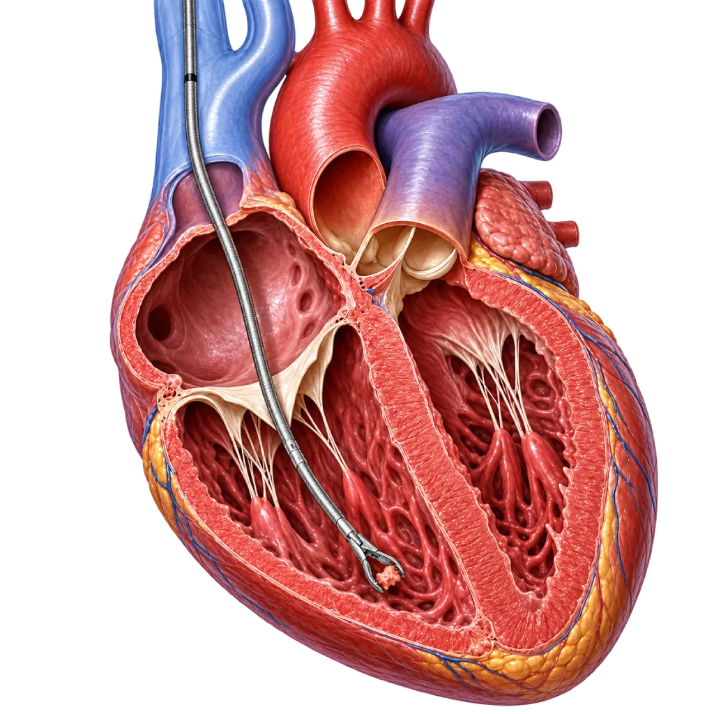
An endomyocardial biopsy is a test that removes several small pieces of heart muscle tissue, each about 1 to 2 millimeters in size. The tissue is taken with a small grasping tool at the tip of a catheter, called a bioptome. This tool is inserted through a vein in the neck or groin into the right ventricle of the heart, and the samples are then examined in a pathology laboratory.
Why Tissue Examination Is Needed
Echocardiography and cardiac MRI can show that the heart muscle is inflamed, thickened, or weakened, but they cannot always confirm the cause. Examining the tissue under a microscope, including special staining (immunohistochemistry) and, if needed, molecular testing, can identify the type of inflammation, a buildup of certain substances such as amyloid, or signs of rejection in a transplanted heart. These results can change treatment choices, for example whether medication that suppresses the immune system needs to be given.
When It Is Usually Done
A biopsy is not performed in every patient with heart muscle disease. Doctors consider it when the result is expected to change management, for example in heart failure that develops quickly and does not improve with treatment, suspected myocarditis with dangerous rhythm disturbances, suspected infiltrative disease that cannot be confirmed in other ways, or routine monitoring after a heart transplant.
Limitations of the Test
Some diseases affect only part of the heart muscle. Because the samples are small, a biopsy may not find an abnormality even when the disease is present. For this reason, biopsy results are always interpreted together with your symptoms, imaging results, and other tests. A biopsy is often performed together with right heart catheterization to measure the pressures inside the heart.
02 / Understanding the procedure
When Is This Procedure Considered?
A doctor may consider this procedure in the situations below. The decision is always made after a thorough assessment.
- Suspected Myocarditis That Is Severe or Not ImprovingEspecially when heart failure develops over days to weeks, is accompanied by dangerous rhythm disturbances or heart block, or does not improve with treatment. Certain types of inflammation require specific treatment that is determined from the tissue results.
- Cardiomyopathy of Unclear CauseWhen heart function declines without a clear cause after a complete workup, a biopsy can help find the cause that determines the direction of treatment.
- Suspected Infiltrative Heart DiseaseSuch as amyloidosis, sarcoidosis, hemochromatosis, or Fabry disease, when the diagnosis cannot yet be confirmed with imaging, blood tests, or a biopsy from another organ.
- Monitoring After a Heart TransplantBiopsies are performed periodically according to the transplant team's schedule to detect signs of organ rejection, often before symptoms appear.
- Heart Damage After Cancer TreatmentIn a small proportion of patients whose heart function declines after chemotherapy, a biopsy is considered when the cause is not yet clear from imaging and other tests.
Situations to Consider First
- Blood Clotting DisordersA severe blood clotting disorder or full-dose blood thinners need to be treated or adjusted beforehand, as directed by your doctor.
- Blood Clots in the Access Route or Inside the HeartA clot in the neck vein, the groin vein, or the right ventricle means the doctor needs to choose another route or postpone the procedure after imaging tests are done.
- Active Infection or FeverAn active infection increases the risk of complications and can affect how the tissue is read, so the procedure is usually postponed until the infection has cleared.
- Results That Would Not Change ManagementWhen the diagnosis can already be confirmed in other ways, or the biopsy result would not change treatment, the risks of the procedure are not justified by its benefits.
03 / Before the procedure
How Should You Prepare?
The heart team will explain the preparation that suits your condition. Patients who are already in the hospital are usually prepared directly by the team on the ward.
Tests before the procedure. Blood tests, including blood clotting and kidney function, an ECG, and echocardiography. Results of a previous cardiac MRI or other tests help the doctor plan the procedure.
Fasting. Follow the fasting instructions from the team, usually for a few hours before the procedure. Routine medications may be taken with a little water if your doctor allows.
Medications you are taking. Tell your doctor about all medications and supplements, especially blood thinners such as warfarin, clopidogrel, or aspirin. Your doctor will decide whether a medication needs to be stopped temporarily and when to restart it. Do not stop any medication on your own.
Allergy history. Tell the team if you have ever been allergic to local anesthetics, contrast dye, or latex.
Documents and clothing. Bring your ID card, referral letter, previous heart test results (ECG, echocardiography, MRI, laboratory), and a list of your medications. Wear loose, comfortable clothing.
Arrival time. Arrive at the time scheduled by the team, ahead of the procedure time, for registration, vital sign checks, and IV line placement.
Consent. Your doctor will explain the purpose, benefits, risks, and alternatives before you sign the consent form for the procedure.
Companion. Arrange for a family member or companion to come with you, because you must not drive yourself after the procedure.
Unsure about the preparation instructions? Ask our team before the procedure.
04 / During the procedure
How Is the Procedure Done?
Endomyocardial biopsy is performed in the catheterization lab (cath lab) by a cardiologist together with nurses and radiographers. You stay awake under local anesthesia and, if needed, are given a mild sedative. The procedure usually takes about half an hour to one hour.
Preparation in the Procedure Room
You lie on the procedure table. ECG electrodes, a blood pressure monitor, and an oxygen level monitor are attached, along with an IV line in your hand.
Local Anesthesia at the Entry Site
The right side of the neck (jugular vein) or the groin (femoral vein) is cleaned and covered with a sterile drape, then injected with a local anesthetic. You will feel a small prick, after which the area becomes numb. The doctor may use ultrasound guidance to locate the vein precisely.
Inserting a Short Tube (Sheath)
A short tube is inserted into the vein as an entry point. You may feel mild pressure, not sharp pain. If needed, the doctor also measures the pressures inside the heart at this stage (right heart catheterization).
Guiding the Bioptome to the Right Ventricle
The bioptome is passed through the tube into the right ventricle of the heart under X-ray guidance, and sometimes echocardiography. Its tip is directed toward the septum, the wall between the two ventricles.
Taking the Tissue Samples
The doctor takes several small pieces of tissue from several spots. When the bioptome touches the heart wall, you may briefly feel a flutter or an irregular heartbeat. This usually goes away on its own within seconds and is closely monitored by the team.
Closing the Entry Site
The bioptome and sheath are removed, then the puncture site is pressed for a few minutes until the bleeding stops and covered with a bandage. You are moved to the recovery room for monitoring, and the samples are sent to the pathology laboratory.
05 / Benefits and risks
Benefits and Risks Should Be Discussed Together.
Every medical procedure has benefits and risks. Your doctor will explain both for your situation before the procedure.
Benefits You Can Expect
- Diagnostic Certainty in Certain SituationsTissue examination can confirm the cause of a heart muscle disease that cannot be confirmed with imaging or blood tests alone.
- Helps Choose TreatmentThe results help the doctor decide on treatment, for example whether medication that suppresses the immune system is needed for certain types of myocarditis.
- Early Detection of Transplant RejectionIn heart transplant patients, signs of organ rejection can be found before symptoms appear, so treatment can be adjusted earlier.
- No Surgical IncisionPerformed through a small puncture in the neck or groin under local anesthesia, without opening the chest.
- Short RecoveryThe majority of patients can return to light activities within one to two days.
The benefits of a procedure are not a promise of a cure. Ask how the results will affect your care plan.
Risks You Should Know
- Bleeding or Bruising at the Puncture SiteThis is common and usually mild. Bleeding usually stops with pressure.
- Heart Rhythm DisturbancesAn irregular rhythm can appear briefly when the bioptome touches the heart wall and usually goes away on its own. A disturbance of the heart's electrical conduction (heart block) is rare and sometimes requires temporary pacing.
- Perforation of the Heart WallA rare but serious complication. Blood can collect in the sac around the heart (tamponade) and must be drained right away with a needle or, very rarely, with surgery.
- Tricuspid Valve InjuryThe bioptome passing through the tricuspid valve can damage the valve and cause it to leak, especially with frequently repeated biopsies. This is rare.
- Air in the Chest Cavity (Pneumothorax)A rare risk with the neck access route. Ultrasound guidance when puncturing the vein helps reduce this risk.
- Accidental Puncture of an Artery Near the VeinAn artery in the neck or groin can also be punctured and cause a large bruise, which is usually managed with pressure.
- InfectionInfection at the puncture site is rare because the procedure is performed using sterile technique.
- Results That Do Not Yet Confirm the DiagnosisBecause the samples are small and the disease may affect only part of the heart muscle, the results may not find an abnormality even though the disease is present. Sometimes other tests or a repeat biopsy are needed.
- Risk of DeathVery rare, and usually related to perforation of the heart wall. Your doctor will explain your estimated risk.
Risks can differ from person to person. A full explanation and consent are given after an individual assessment.
06 / After the procedure
What Is Recovery Like?
Recovery after an endomyocardial biopsy is usually quick, but the underlying heart condition also affects how long you need to stay in care.
The initial few hours. Your blood pressure, heart rhythm, and puncture site are checked regularly. The doctor may perform echocardiography to make sure there is no fluid around the heart. If the puncture was in the groin, you will be asked to lie with your leg straight for a few hours.
Hospital stay. Some patients can go home the same day or the next day. Patients who are already in the hospital for their heart condition continue their care according to the doctor's plan.
The next one to two days. Mild pain or bruising at the puncture site is normal. Keep the area clean and dry, and avoid strenuous activity. Light activities can usually be resumed within one to two days, as advised by your doctor.
Test results. Tissue examination in the laboratory takes time, usually about one week, and longer if special staining or molecular testing is needed. Your doctor will schedule a consultation to explain the results and the treatment plan.
Warning signs. Contact the medical team right away or go to the emergency room if you experience:
- Bleeding from the puncture site that does not stop after a few minutes of pressure, or a lump that grows quickly
- Chest pain, sudden shortness of breath, or dizziness as if you are about to faint
- A very fast or irregular heartbeat
- Fever, or a puncture site that becomes red, swollen, and filled with pus
- Numbness, coldness, or severe pain in the leg used as the access route
Know the Signs That Need Prompt Care.
Follow the team's guidance on warning signs. If severe or sudden symptoms appear after the procedure, go to the nearest emergency department right away or call Heart Attack Center 1500 258.
07 / Team and support
Doctors and Procedure Support
The doctors below are listed for this procedure. Listed by name, not by ranking.

Dr. dr. Dafsah A. Juzar, Sp.JP(K)
Interventional Cardiology
Coronary Intervention Procedures +1 center
Clinical focusComplex Coronary Intervention · Cardiac Catheterization
View Profile
Dr. dr. Jajang Sinardja, Sp.JP(K)
Interventional Cardiology
Coronary Intervention Procedures
Clinical focusAcute Coronary Syndrome · Primary Coronary Intervention
View Profile
dr. Siska S. Danny, Sp.JP(K)
Interventional Cardiology
Coronary Intervention Procedures
Clinical focusPCI for Acute Coronary Syndrome · Primary Coronary Intervention
View Profile08 / Common questions
Before You Plan the Procedure.
Questions that patients and families often ask before an endomyocardial biopsy. Answers specific to your situation are given by your doctor during a consultation.
Does a heart biopsy hurt?
The procedure is performed under local anesthesia, so the puncture site feels numb. You may feel pressure when the tube is inserted and a brief flutter when the samples are taken. If needed, the doctor gives a mild sedative to help you relax. After the procedure, mild pain at the puncture site can usually be relieved with ordinary pain relievers.
Will my heart become weaker after tissue is taken from it?
No. The pieces of tissue taken are very small, each about 1 to 2 millimeters, from the thick, muscular wall of the heart. The sampled spots heal on their own and do not affect the heart's pumping strength. The main risks of this procedure are rare complications, such as perforation of the heart wall, which the team monitors closely during and after the procedure.
How long does it take to get the biopsy results?
Tissue examination in the pathology laboratory usually takes about one week. If special staining, molecular testing, or testing at a referral laboratory is needed, it may take longer. While you wait, continue your medications as prescribed. Your doctor will schedule a consultation to explain the results and your treatment plan.
Why do I still need a biopsy if I have already had a cardiac MRI?
A cardiac MRI can show inflammation, scar tissue, or a buildup of substances in the heart muscle, but it cannot always confirm the specific cause. In certain situations, such as suspected myocarditis of a particular type or infiltrative disease, the type of cause determines treatment. Tissue examination helps the doctor obtain that certainty.
Are biopsy results always conclusive?
Not always. Some diseases affect only part of the heart muscle, so a small sample may miss the affected area. For this reason, the doctor takes several samples and interprets the results together with your symptoms, imaging, and other tests. If the results do not yet answer the clinical question, the doctor may recommend other tests or a repeat biopsy.
Does the biopsy need to be repeated?
In heart transplant patients, biopsies are performed periodically according to the transplant team's schedule, especially during the initial year after the transplant, when the risk of rejection is still high. In other situations, one biopsy is usually enough, unless the results have not confirmed the diagnosis or the doctor needs to assess the response to ongoing treatment.
Sources and Medical Review
Translated from the Indonesian edition medically reviewed by dr. Sri Diniharini, Sp.JP(K).
Sources as cited in the Indonesian edition.
- Cooper LT, Baughman KL, Feldman AM, et al. The Role of Endomyocardial Biopsy in the Management of Cardiovascular Disease: A Scientific Statement From the American Heart Association, the American College of Cardiology, and the European Society of Cardiology. Circulation. 2007;116(19):2216-2233.
- Caforio ALP, Pankuweit S, Arbustini E, et al. Current state of knowledge on aetiology, diagnosis, management, and therapy of myocarditis: a position statement of the ESC Working Group on Myocardial and Pericardial Diseases. Eur Heart J. 2013;34(33):2636-2648.
- Arbelo E, Protonotarios A, Gimeno JR, et al. 2023 ESC Guidelines for the management of cardiomyopathies. Eur Heart J. 2023;44(37):3503-3626.
- Drazner MH, Bozkurt B, Cooper LT, et al. 2024 ACC Expert Consensus Decision Pathway on Strategies and Criteria for the Diagnosis and Management of Myocarditis. J Am Coll Cardiol. 2025;85(4):391-431.
- Stewart S, Winters GL, Fishbein MC, et al. Revision of the 1990 working formulation for the standardization of nomenclature in the diagnosis of heart rejection. J Heart Lung Transplant. 2005;24(11):1710-1720.