
Quick answer
What Is Hypoplastic Left Heart Syndrome?
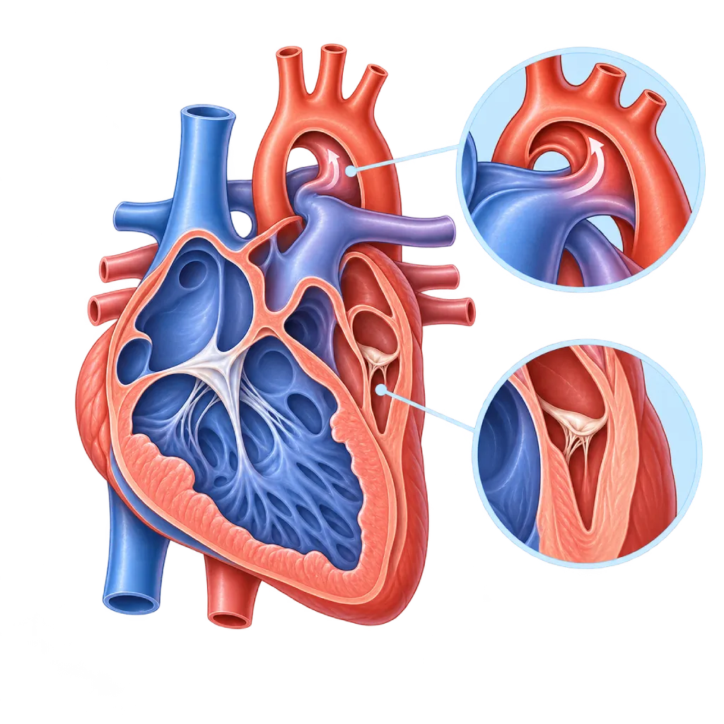
Hypoplastic left heart syndrome is a severe congenital heart defect in which the left ventricle, mitral valve, aortic valve, and aorta are very small. The left side of the heart cannot pump blood to the body, so the baby needs urgent care and staged surgery.
Sources01 / Understanding the condition
About Hypoplastic Left Heart Syndrome
In a normal heart, the right side pumps blood to the lungs to pick up oxygen, while the left side pumps oxygen-rich blood to the whole body. In hypoplastic left heart syndrome (known by the abbreviation HLHS), the left side does not grow properly during pregnancy. The left ventricle is very small, the mitral and aortic valves are narrowed or closed, and the base of the aorta is small. This condition is a critical congenital heart disease and occurs in about 2 to 3 of every 10,000 births, more often in baby boys. Other types of congenital heart defects are covered on the Congenital Heart Disease page.
Why Babies Can Get Worse After Birth
In the womb, the baby's blood passes through two natural shortcuts: the foramen ovale in the wall between the upper chambers, and the ductus arteriosus between the pulmonary artery and the aorta. After birth, babies with HLHS depend on both of these pathways. The right ventricle pumps blood to the lungs and, through the ductus, to the whole body. The ductus usually starts to close in the first few days. When that happens, blood flow to the body drops sharply and the baby can go into shock. This is why a baby can look well in the first hours and then get worse quickly.
Staged Treatment
The left side of the heart cannot be made to grow again. The goal of treatment is to make the right ventricle work as the single pump for the body. Usually three stages of surgery are done: the Norwood procedure or a hybrid approach in the first weeks, the Glenn operation at about 3 to 6 months of age, and the Fontan operation at about 2 to 5 years of age. For some babies, a heart transplant is an option. Families also have the right to a full explanation of all options, including care that focuses on the baby's comfort.
Life After Surgery
Many children with HLHS now grow up to school age and adulthood. Even so, a circulation with a single ventricle, called the Fontan circulation, needs lifelong monitoring of pump function, the tricuspid valve, heart rhythm, the liver, and the child's growth and development. The 2019 AHA scientific statement and the 2020 ESC guidelines recommend regular checkups at a service experienced in congenital heart disease.
02 / Understanding the condition
Symptoms to Recognize.
Common Symptoms
- Skin, lips, or nails that look bluish or grayish
- Fast breathing or breathing that looks like hard work
- Difficulty feeding and tiring quickly while feeding
- Cold hands and feet, with a weak pulse
- A baby who seems sluggish or very sleepy
- Low oxygen saturation on newborn screening
Symptoms in Newborns
A baby may look well in the first hours. Symptoms often appear in the first few days, when the ductus arteriosus starts to close.
- Skin color turning pale gray
- Breathing that gets faster over one to two days
- Refusing to feed or falling asleep while feeding
- Fewer wet diapers
- A body that feels cold and clammy
Symptoms in Infants
The period between the first and second stage operations needs close monitoring at home as directed by the team.
- Weight not increasing as planned
- Oxygen saturation below the limit set by the doctor
- Looking bluer than usual
- Vomiting, diarrhea, or difficulty drinking
- Faster breathing or sweating while feeding
- Unusual fussiness or more sluggishness than usual
Symptoms in Children
After the Fontan operation, symptoms are usually mild, but they should be reported at checkups.
- Tiring more quickly than peers
- Slightly bluish lips during activity
- A swollen abdomen or swelling in the legs
- Persistent diarrhea or weight loss
- Palpitations or a past fainting episode
- Slower growth or development
03 / Understanding the condition
What Causes This Condition?
- Abnormal growth of the left side of the baby's heart in early pregnancy, the exact cause of which is often unknown
- Very low blood flow through the baby's mitral or aortic valve before birth, so the left ventricle does not grow
- Genetic changes in some babies, including chromosome abnormalities such as Turner syndrome
- A combination of genetic and environmental factors during pregnancy
04 / Understanding the condition
Who Is at Higher Risk?
Genetic
A history of hypoplastic left heart syndrome or another left-sided heart defect, such as a bicuspid aortic valve, in a parent or sibling increases the risk in future pregnancies.
Medical
Uncontrolled diabetes before and during pregnancy, as well as some infections and medicines in the first trimester, are linked to a higher risk of congenital heart disease.
Lifestyle
Smoking and drinking alcohol during pregnancy increase the risk of congenital heart defects in general, although the link to this syndrome is not specific.
Other
This condition is more common in baby boys. In a pregnancy without a targeted ultrasound, the defect may only be discovered after the baby is born and suddenly gets worse.
Groups at Higher Risk
- Unborn Babies with a Suspected Heart Defect: A left side of the heart that looks small on a pregnancy ultrasound needs to be confirmed with fetal echocardiography so the delivery can be planned.
- Families with a History of Left Heart Defects: A parent or sibling with a left-sided heart defect adds to the risk, so fetal echocardiography is recommended in future pregnancies.
- Babies Between the Norwood and Glenn Operations: The period between the first and second stage operations requires daily monitoring of weight, oxygen saturation, and intake.
- Teenagers and Adults with a Fontan Circulation: Survivors are at risk of rhythm problems, liver problems, fluid buildup, and declining pump function over time.
05 / Understanding the condition
How Is It Diagnosed?
Many cases can already be suspected on a pregnancy ultrasound, when the left side of the baby's heart looks small. This suspicion is confirmed with fetal echocardiography. A diagnosis before birth gives time to plan the delivery at a hospital able to care for babies with critical heart disease, and to start medicine right after the baby is born.
If it was not detected before birth, early clues may include low oxygen saturation on newborn screening, fast breathing, a weak pulse, or a baby who suddenly becomes sluggish. Echocardiography confirms the diagnosis by assessing the size of the left ventricle, the mitral and aortic valves, the aortic arch, right ventricle function, the tricuspid valve, and the opening in the wall between the upper chambers. A chest X-ray, an ECG, and blood tests such as blood gases and kidney function help assess how serious the baby's condition is. Cardiac catheterization is done when pressures need to be measured or when an opening between the upper chambers that is too narrow needs to be widened.
Fetal echocardiography: a focused ultrasound of the baby's heart, usually at 18 to 22 weeks of pregnancy, to confirm a suspicion from a routine ultrasound.
Pulse oximetry screening: sensors on the right hand and a foot of the newborn to detect low oxygen levels before symptoms are obvious.
Echocardiography
The main test after birth to confirm the size of the left ventricle, the valves, the aorta, right ventricle function, the tricuspid valve, and the opening in the wall between the upper chambers.
Chest X-Ray
Assesses heart size and blood flow in the lungs, and the position of tubes and catheters while the baby is in intensive care.
Electrocardiogram (ECG)
Records the rhythm and the strain on the heart chambers, both at diagnosis and during monitoring after staged surgery.
Cardiac Catheterization
Measures pressure in the lungs before the Glenn and Fontan operations, and can widen an opening between the upper chambers that is too narrow.
Cardiac MRI
Used in children and adults after the Fontan operation to assess pump function, blood flow, and the blood vessels around the heart.
06 / Understanding the condition
Treatment Options.
Treatment is decided by a doctor after an assessment. The categories below can help you follow the conversation with your doctor.
Stabilization in the Intensive Care Unit
Prostaglandin medicines given through an IV keep the ductus open. Oxygen and breathing support are carefully adjusted to keep blood flow to the lungs and to the body balanced.
Norwood Operation
The first stage, in the first weeks: the base of the pulmonary artery and the small aorta are joined to form a new aorta, then blood flow to the lungs is provided through a small tube (shunt).
Hybrid Approach
In certain babies, the first stage is done by placing a stent in the ductus and narrowing the pulmonary arteries, combining surgery and catheter techniques. Covered on the Hybrid Heart Procedure page.
Glenn Operation
The second stage, at about 3 to 6 months of age: the large vein from the upper body is connected directly to the pulmonary artery.
Fontan Operation
The third stage, at about 2 to 5 years of age: blood from the lower body is also routed directly to the lungs, so the right ventricle pumps only to the body.
Heart Transplant
Considered when staged surgery is not suitable or heart function declines later in life. Covered on the Heart Transplant page.
Comfort-Focused Care
Some families, after a full discussion with the team, choose care that puts the baby's comfort first. This choice is respected and supported.
07 / Understanding the condition
Complications to Watch For.
Shock in Newborns
When the ductus arteriosus closes, blood flow to the body can drop suddenly and affect the kidneys, intestines, liver, and brain.
Reduced Right Ventricle Function
The right ventricle is not designed to pump blood to the whole body for a lifetime, so its strength can decline over time.
Leaking Tricuspid Valve
The valve between the right upper and lower chambers can leak and add to the pump's workload. Some cases need surgical repair.
Heart Rhythm Disorders
Arrhythmias can develop after staged surgery, especially after the Fontan operation. Covered on the Heart Rhythm Disorders / Arrhythmia page.
Fontan Circulation Complications
Long-term high pressure in the veins can affect the liver, cause protein to leak through the intestines, and make blood clots more likely to form.
Delayed Growth and Development
Some children have delays in movement, speech, or learning, so developmental monitoring is recommended from an early age.
08 / Understanding the condition
What Can You Do?
- There is no sure way to prevent this condition yet, but health before and during pregnancy is still important
- Control diabetes before pregnancy, take folic acid as recommended, and avoid smoking and alcohol
- Have scheduled pregnancy ultrasounds, and fetal echocardiography if your doctor finds something suspicious
- If there is a family history, tell your obstetrician so the baby's heart can be checked more carefully
- If the condition has been diagnosed, plan the delivery at a hospital able to care for babies with critical heart disease
- Record the baby's weight, intake, and oxygen saturation every day between operations as directed by the team
- Keep your child's immunizations up to date and look after their teeth to reduce the risk of infection
- Follow the lifelong checkup schedule after the Fontan operation even if your child looks healthy
09 / Understanding the condition
When Should You See a Doctor?
Take your baby to the emergency room right away if they look pale gray or increasingly blue, are very limp, are hard to wake, breathe very fast, or refuse to feed. This condition can get worse within hours. Talk to a doctor promptly if:
- A pregnancy ultrasound shows that the left side of the baby's heart is small, or shows another heart abnormality.
- Your baby's oxygen saturation screening result is abnormal.
- Between operations, your baby's weight is not increasing, saturation drops below the limit, or your baby has vomiting and diarrhea.
- Your child has swelling, a swollen abdomen, persistent diarrhea, palpitations, or fainting after the Fontan operation.
- A teenager or adult with a Fontan circulation has not had regular checkups.
10 / Common questions
Questions You May Have.
Frequently asked questions about hypoplastic left heart syndrome.
Can hypoplastic left heart syndrome be seen during pregnancy?
Often, yes. A small left side of the heart usually shows up on a pregnancy ultrasound in the second trimester and is then confirmed with fetal echocardiography. A diagnosis before birth helps the family and the team plan where the baby will be delivered, prepare intensive care, and start medicine right after birth. Even so, some cases are only found after birth, so newborn oxygen saturation screening is still important.
Why does the surgery have to be done in stages?
Pressure in a newborn's lungs is still high, so blood cannot yet flow to the lungs without being pumped. That is why the first operation creates a new aorta and a blood path to the lungs through a small tube. After lung pressure falls and the baby grows, the Glenn and then the Fontan operation route blood from the veins directly to the lungs. These stages give the child's body time to adjust to a single-ventricle circulation.
What should we watch for at home after the first stage operation?
The period between the Norwood and Glenn operations needs close monitoring. The team usually asks families to weigh the baby, record how much the baby drinks, and measure oxygen saturation every day. Report right away if saturation drops below the set limit, weight is not increasing, or the baby has vomiting, diarrhea, or fever, or looks bluer and more sluggish. In studies, home monitoring programs like this have been linked to better outcomes.
Can a child with a Fontan circulation go to school and play as usual?
Many children with a Fontan circulation go to school, play, and do light to moderate exercise. Their physical capacity is usually lower than that of their peers, and they need to drink enough fluids, especially in hot weather or when sick. Regular physical activity is actually encouraged to maintain fitness. Your doctor will tailor the types of safe exercise based on your child's heart function, rhythm, and oxygen saturation.
Are future pregnancies also at risk?
The chance of a heart defect in a future pregnancy is higher than in the general population, although the majority of babies born afterward have a normal heart. The defect that appears is not always the same, for example a bicuspid aortic valve. That is why fetal echocardiography is recommended in future pregnancies. Genetic counseling can help families understand this chance, especially if there are other abnormalities outside the heart or a family history.
Sources and Medical Review
Translated from the Indonesian edition medically reviewed by dr. Radityo Prakoso, Sp.JP(K).
Sources as cited in the Indonesian edition.
- Feinstein JA, Benson DW, Dubin AM, et al. Hypoplastic left heart syndrome: current considerations and expectations. J Am Coll Cardiol. 2012;59(1 Suppl):S1-S42.
- Ohye RG, Sleeper LA, Mahony L, et al. Comparison of shunt types in the Norwood procedure for single-ventricle lesions. N Engl J Med. 2010;362(21):1980-1992.
- Newburger JW, Sleeper LA, Gaynor JW, et al. Transplant-Free Survival and Interventions at 6 Years in the SVR Trial. Circulation. 2018;137(21):2246-2253.
- Rychik J, Atz AM, Celermajer DS, et al. Evaluation and Management of the Child and Adult With Fontan Circulation: A Scientific Statement From the American Heart Association. Circulation. 2019;140(6):e234-e284.
- Donofrio MT, Moon-Grady AJ, Hornberger LK, et al. Diagnosis and treatment of fetal cardiac disease: a scientific statement from the American Heart Association. Circulation. 2014;129(21):2183-2242.
- Martin GR, Ewer AK, Gaviglio A, et al. Updated Strategies for Pulse Oximetry Screening for Critical Congenital Heart Disease. Pediatrics. 2020;146(1):e20191650.
- Baumgartner H, De Backer J, Babu-Narayan SV, et al. 2020 ESC Guidelines for the management of adult congenital heart disease. Eur Heart J. 2021;42(6):563-645.
Doctors Who Treat This Condition

dr. Radityo Prakoso, Sp.JP(K)
Pediatrics & Congenital Heart Disease
Congenital and Structural Heart Center
Clinical focusPediatric Cardiology Consultation · Congenital Heart Disease Care
View Profile
dr. Salomo Purba, Sp.BTKV, Subsp.JPK(K)
Pediatric and Congenital Heart Surgery
Congenital and Structural Heart Center +2 centers
Clinical focusPediatric Heart Surgery · Congenital Heart Defect Surgery
View Profile
dr. Tartila, Sp.A, Subsp.ETIA(K)
Pediatric Emergency and Intensive Care
Congenital and Structural Heart Center
Clinical focusPediatric Intensive Care · Pediatric Emergency Care
View Profile