
Quick answer
What Is Peripheral Angiography?
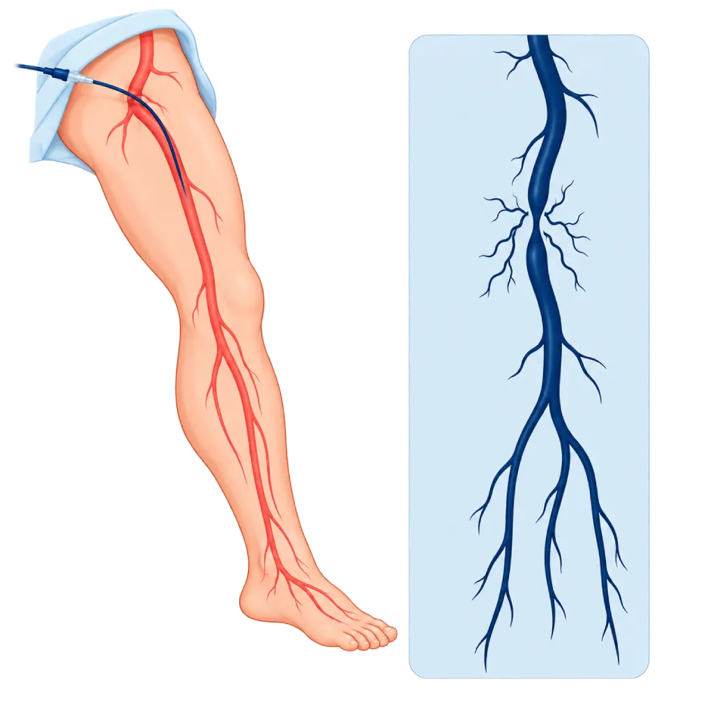
Peripheral angiography is an examination of the leg or arm arteries using a thin catheter and contrast dye recorded by X-ray. It shows the location, length, and severity of narrowing in detail, down to the arteries of the foot. If needed, the narrowing can be opened right away with a balloon or stent in the same session.
Sources01 / Understanding the procedure
About Peripheral Angiography
What Is Peripheral Angiography
Peripheral angiography examines the arteries outside the heart, mainly the leg arteries and sometimes the arm arteries. A thin catheter is inserted into an artery, then contrast dye is injected so the arteries show up clearly on X-ray images. This examination provides a very detailed map of the arteries, so it is often done when a procedure to open a narrowing is being planned.
How It Works in Brief
The catheter is inserted through an artery in the groin or wrist, then contrast dye is injected. X-rays record the flowing contrast dye, and a computer removes the image of the bones so only the blood vessels are visible.
What Can Be Seen
- Narrowing and blockages: their location, length, and severity.
- Collateral vessels: small side pathways that form to carry blood around a blockage.
- Arteries below the knee and in the foot: important for assessing foot wounds that are slow to heal.
- Other abnormalities: for example a widened artery (aneurysm) or a clot causing a sudden blockage.
- Previous stents or bypasses: to see whether they are still open.
Diagnosis and Treatment in One Session
If a suitable narrowing is found, the doctor can widen it right away with a balloon or place a stent through the same catheter, without a second puncture. This possibility is discussed and agreed on with you before the examination.
Catheter Angiography, CT, or MR Angiography
CT angiography and MR angiography also show the arteries without inserting a catheter into an artery. Catheter angiography is usually chosen when a procedure is planned, when the results of other tests are unclear, or to assess the small arteries below the knee.
02 / Understanding the procedure
When Is This Procedure Considered?
A doctor may consider this procedure in the situations below. The decision is always made after a thorough assessment.
- Planning a Balloon, Stent, or Bypass SurgeryProvides a detailed map of the arteries before or during a procedure to restore blood flow.
- Slow-Healing Foot Wounds or Pain at RestTo view the arteries down to the foot and find a way to restore blood flow.
- Limiting Leg Pain When WalkingWhen medicines and walking exercise have not helped enough and a procedure is being considered.
- Unclear Results from Other TestsWhen duplex ultrasound, CT, or MR angiography has not adequately answered the doctor's questions.
- Sudden Blockage in a Leg or ArmWhen a clot is suspected of blocking an artery, to decide on treatment and sometimes to carry it out right away.
Situations to Consider First
- Severely Impaired Kidney FunctionContrast dye can worsen kidney problems. The doctor weighs the benefits, gives IV fluids, and limits the amount of contrast dye.
- Allergy to Contrast DyeAllergy-preventing medicine can be given beforehand. Tell the team from the start.
- Blood Clotting DisordersThe risk of bleeding at the puncture site increases, so clotting is checked and medicines are adjusted beforehand.
- PregnancyX-ray examinations are avoided during pregnancy unless truly necessary.
- Infection or a Wound at the Puncture SiteThe catheter is inserted at another location, for example the wrist or the groin on the other side.
03 / Before the procedure
How Should You Prepare?
The team will give you written instructions before the examination.
Blood tests and previous results. Kidney function and blood clotting are checked beforehand. Bring the results of a duplex ultrasound, ABI test, CT angiography, or records of previous blood vessel procedures.
Medicines you are taking. Bring a list of all your medicines and supplements. Aspirin and antiplatelet medicines are usually continued, while other blood thinners and some diabetes medicines such as metformin may need to be adjusted. Do not stop any medicine without your doctor's direction.
Fasting. Usually for a few hours before the examination, according to the team's instructions.
Allergies. Tell the team if you have ever been allergic to contrast dye, medicines, adhesive tape, or latex.
Pregnancy. Tell the team if you are or might be pregnant.
Companion. Arrange for someone to take you home, because you are advised not to drive on the day of the examination.
Unsure about the preparation instructions? Ask our team before the procedure.
04 / During the procedure
How Is the Procedure Done?
The examination usually takes about one hour. It takes longer if it is followed right away by balloon or stent placement.
Preparation in the Procedure Room
You lie on the examination table, an IV line is placed, and your heart and blood pressure are monitored. A mild sedative may be given if you feel anxious.
Blood Vessel Access
After local anesthesia, the doctor inserts a small tube into an artery, usually in the groin and sometimes in the wrist. You stay awake and feel only pressure.
Taking the Images
The catheter is guided to the artery being examined, then contrast dye is injected while X-rays record the images. You may feel warmth in your leg for a few seconds and will be asked to stay still for a moment.
Further Treatment When Needed
If a suitable narrowing is found and treatment has been agreed on beforehand, the doctor can widen it right away with a balloon or place a stent.
Closing the Puncture Site
The tube is removed, then the puncture site is pressed or closed with a special device to prevent bleeding. After that, you are monitored in the recovery room.
05 / Benefits and risks
Benefits and Risks Should Be Discussed Together.
Every medical procedure has benefits and risks. Your doctor will explain both for your situation before the procedure.
Benefits You Can Expect
- A Detailed View of the ArteriesShows the arteries down to the foot, including small vessels below the knee.
- Diagnosis and Treatment at OnceNarrowing can be opened right away with a balloon or stent without a second puncture.
- A Basis for Choosing TreatmentHelps decide between medicines, a catheter procedure, or bypass surgery.
- Helping Save the LimbIn foot wounds or severely reduced blood flow, it shows the pathways that can be opened.
- No Large IncisionOnly a small puncture is needed, and the majority of patients can go home on the same day.
The benefits of a procedure are not a promise of a cure. Ask how the results will affect your care plan.
Risks You Should Know
- Bleeding or Bruising at the Puncture SiteCommon and usually mild. Sometimes a lump forms that needs additional treatment.
- Reaction to Contrast DyeRanging from rash and itching to, very rarely, a severe reaction that the team treats right away.
- Kidney Function ProblemsEspecially if kidney function is already reduced or you have diabetes. Usually temporary.
- Blood Vessel InjuryVery rare, for example a tear or a clot in the artery the catheter passes through.
- X-Ray ExposureThe dose is kept as low as possible for the needs of the examination.
Risks can differ from person to person. A full explanation and consent are given after an individual assessment.
06 / After the procedure
What Is Recovery Like?
If only the examination is done, the majority of patients can go home on the same day after a few hours of monitoring. If it is followed by a balloon or stent, some patients stay in the hospital for one night.
After the examination. If the catheter was inserted through the groin, you lie down for a few hours with your leg straight. If it was inserted through the wrist, a compression band is worn for a few hours.
Drink water. Drink enough water to help your kidneys clear the contrast dye, unless your doctor limits your fluids.
Activity. Avoid lifting heavy objects and strenuous exercise for a few days, and do not drive on the day of the examination.
Medicines. If a stent was placed, take your antiplatelet medicine every day as prescribed and do not stop it on your own.
Results. The doctor explains the findings and the next steps, for example medical treatment, further procedures, or bypass surgery.
Warning signs. Go to the emergency room right away or contact the medical team if you experience:
- Swelling that gets bigger, or bleeding, at the puncture site
- A leg or arm that feels cold, pale, numb, or painful
- Rash, itching, or shortness of breath after the examination
- A much smaller amount of urine than usual
Know the Signs That Need Prompt Care.
Follow the team's guidance on warning signs. If severe or sudden symptoms appear after the procedure, go to the nearest emergency department right away or call Heart Attack Center 1500 258.
07 / Team and support
Doctors and Procedure Support
The doctors below are listed for this procedure. Listed by name, not by ranking.

dr. Emanoel Oepangat, Sp.JP(K)
Interventional Cardiology
Vascular Diagnostic and Therapeutic Center +1 center
Clinical focusCoronary Intervention · Peripheral Vascular Intervention
View Profile
dr. Suko Adiarto, Sp.JP(K), Ph.D.
Interventional Cardiology
Vascular Diagnostic and Therapeutic Center +1 center
Clinical focusCoronary Intervention · Peripheral Vascular Intervention
View ProfileVascular Interventional Suite
A procedure room with X-ray imaging for diagnosing and treating narrowed, blocked, or enlarged blood vessels outside the heart…
The availability of technology and test support is confirmed with the team. Technology is chosen according to clinical need.
08 / Common questions
Before You Plan the Procedure.
Frequently asked questions about peripheral angiography. Decisions about the examination and any further procedures are made together with your doctor.
What is the difference between peripheral angiography and CT angiography?
CT angiography images the arteries with contrast dye injected through an IV line in the arm, without a catheter inside the artery. Peripheral angiography places a catheter in the artery, so the images are more detailed and narrowing can be treated right away. That is why catheter angiography is usually chosen when a procedure is planned or when the results of other tests are unclear.
Is peripheral angiography painful?
The puncture site is numbed with a local anesthetic, so you only feel pressure when the tube is inserted. When the contrast dye is injected, your leg may feel warm for a few seconds. The catheter moving inside the artery cannot be felt. A mild sedative can be given if you feel anxious, and you can tell the team at any time if you feel uncomfortable.
Can I go home on the same day?
If only the examination is done, the majority of patients can go home after a few hours of monitoring, as long as the puncture site is secure. If it is followed by balloon or stent placement, some patients need to stay in the hospital for one night. Arrange for someone to take you home, because you are advised not to drive on the day of the examination.
Can a narrowing be opened right away during angiography?
Yes, if the narrowing is suitable for a catheter procedure and this has been agreed on beforehand. The doctor can widen it with a balloon or place a stent through the same catheter. If the blockage is too long or complex, the doctor will discuss other options, such as bypass surgery, after the examination.
What if my kidney function is reduced?
Contrast dye can affect the kidneys, especially if their function is already reduced. The doctor weighs the benefits of the examination, gives IV fluids before and after, and uses as little contrast dye as possible. Kidney function can be rechecked after the examination. Tell the team if you undergo regular dialysis.
How long does the examination take?
The examination usually takes about one hour, not including preparation and monitoring afterward. If it is followed right away by a balloon or stent, it takes longer. Overall, plan to spend half a day or more at the hospital, including lying down afterward, and follow the time estimate given by the team.
Sources and Medical Review
Translated from the Indonesian edition medically reviewed by dr. Emanoel Oepangat, Sp.JP(K).
Sources as cited in the Indonesian edition.
- Mazzolai L, Teixido-Tura G, Lanzi S, et al. 2024 ESC Guidelines for the management of peripheral arterial and aortic diseases. Eur Heart J. 2024;45(36):3538-3700.
- Gornik HL, Aronow HD, Goodney PP, et al. 2024 ACC/AHA/AACVPR/APMA/ABC/SCAI/SVM/SVN/SVS/SIR/VESS Guideline for the Management of Lower Extremity Peripheral Artery Disease. Circulation. 2024;149(24):e1313-e1410.
- Conte MS, Bradbury AW, Kolh P, et al. Global vascular guidelines on the management of chronic limb-threatening ischemia. J Vasc Surg. 2019;69(6S):3S-125S.e40.
- van der Molen AJ, Reimer P, Dekkers IA, et al. Post-contrast acute kidney injury. Part 1: Definition, clinical features, incidence, role of contrast medium and risk factors. Eur Radiol. 2018;28(7):2845-2855.