
Quick answer
What Is Peripheral CT Angiography?
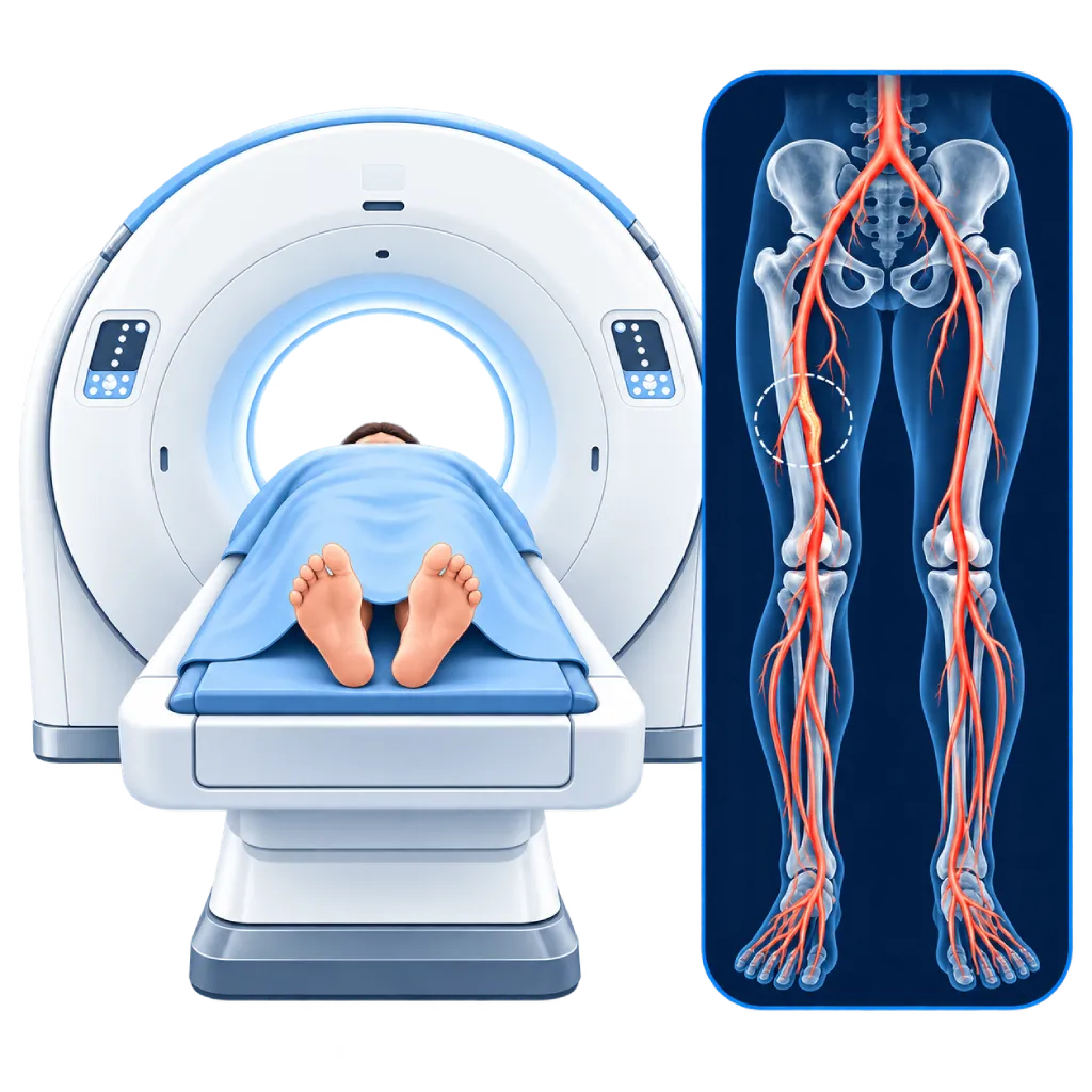
Peripheral CT angiography (peripheral CTA) examines the leg arteries using a CT scanner and contrast dye injected through an IV in your arm. The scan takes only a few seconds and creates three-dimensional images of the arteries from the abdomen to the feet, showing the doctor the location and severity of narrowing without a catheter in the artery.
Sources01 / Understanding the procedure
About Peripheral CT Angiography
What Is Peripheral CT Angiography
Peripheral CT angiography, or peripheral CTA, examines the arteries that carry blood to the legs, from the aorta in the abdomen through the pelvic and thigh arteries down to the feet. This test uses a CT scanner, which is an X-ray beam that rotates around the body, together with an iodine-based contrast dye that makes the arteries clearly visible. No catheter is inserted into the arteries.
How It Works in Brief
You lie on a table that moves through the ring of the CT scanner. Contrast dye is injected through an IV in your arm, then the scanner scans from the abdomen to the feet within seconds as the contrast fills the arteries. A computer then builds three-dimensional images of the arteries along with the surrounding bones.
What Can Be Seen
- Narrowings and blockages: their location, length, and severity, from the abdomen to the feet.
- Calcium buildup in the artery walls: important for planning a balloon, stent, or surgery.
- Previous stents or bypasses: to see whether they are still open.
- Widened arteries: for example an abdominal aortic aneurysm or an aneurysm of the artery behind the knee.
- Access routes for procedures: helps choose the artery for catheter entry if a procedure is planned.
CTA, MR Angiography, or Catheter Angiography
Duplex ultrasound is usually the initial test. If a more complete map of the arteries is needed, CTA is often chosen because it is fast and widely available, including for people with a pacemaker. MR angiography may be considered if X-rays or iodine-based contrast need to be avoided, or if calcium buildup in the arteries is very heavy. Catheter angiography is usually chosen when a procedure to open the narrowing is already planned.
02 / Understanding the procedure
When Is This Procedure Considered?
A doctor may consider this procedure in the situations below. The decision is always made after a thorough assessment.
- Limiting Leg Pain When WalkingWhen a procedure is being considered and the doctor needs a complete map of the leg arteries.
- Slow-Healing Foot Wounds or Pain at RestTo map the arteries down to the feet and find a route to restore blood flow.
- Planning a Balloon, Stent, or Bypass SurgeryShows the location of narrowings, calcium buildup, and the routes that can be used for the procedure.
- Checking a Previous Stent or BypassIf symptoms come back after a procedure, to see whether the stent or bypass is still open.
- Widening of an Artery or the AortaWhen an abdominal aortic aneurysm or a leg artery aneurysm is suspected.
- Sudden Blockage in the LegThe test is fast, which helps the doctor decide on treatment quickly.
Situations to Consider First
- Severe Allergy to Iodine-Based ContrastThe doctor will consider other tests, or give allergy-prevention medication beforehand.
- Severe Kidney ImpairmentThe doctor weighs the benefits, gives IV fluids if needed, or chooses another test.
- PregnancyX-ray tests are avoided during pregnancy unless truly necessary.
- Uncontrolled HyperthyroidismThe iodine in the contrast can worsen the thyroid condition, so the timing of the test is discussed with your doctor.
- Difficulty Lying StillMovement blurs the images. A mild sedative can help if you feel anxious.
03 / Before the procedure
How Should You Prepare?
The team will give you written instructions before the test.
Kidney function. If you have kidney disease or diabetes, or are of advanced age, your kidney function is checked beforehand. Bring your recent blood test results if you have them.
Current medications. Bring a list of all your medications. Regular medications are usually continued. If your kidney function is severely reduced, metformin may need to be stopped temporarily. Do not stop any medication without your doctor's guidance.
Eating and drinking. Some hospitals ask you not to eat for a few hours beforehand. Follow the team's instructions. Drinking water is usually still allowed.
Allergies and thyroid. Tell the team if you have ever had an allergy to contrast dye or have a thyroid disorder.
Pregnancy. Tell the team if you are or might be pregnant.
Clothing. Wear comfortable clothing, and remove belts, jewelry, and metal objects around the abdomen and legs.
Unsure about the preparation instructions? Ask our team before the procedure.
04 / During the procedure
How Is the Procedure Done?
The time in the CT room is usually about 15 to 30 minutes, while the scan itself takes only a few seconds.
Preparation in the CT Room
An IV line is placed in your arm for the contrast. You lie on your back on the exam table, usually with your feet toward the scanner. Both legs are positioned straight and supported so they do not move.
Initial Images
The scanner takes initial images to set the scan range from the abdomen to the feet. The staff talk with you through an intercom and give you instructions.
Contrast Injection and Scanning
The contrast is injected through the IV with a pump. You may feel warmth throughout your body or feel as if you need to urinate for a few seconds, and this is normal. You are asked to hold your breath briefly and stay still while the table moves through the ring of the scanner.
Completing the Test
You may be asked to wait for a short time to make sure there is no reaction to the contrast, then the IV is removed. The images are processed into a three-dimensional map of the arteries, then analyzed and reported by a doctor.
05 / Benefits and risks
Benefits and Risks Should Be Discussed Together.
Every medical procedure has benefits and risks. Your doctor will explain both for your situation before the procedure.
Benefits You Can Expect
- FastThe scan takes only a few seconds, so it is also suitable for urgent situations.
- No Catheter in the ArteryThe contrast is simply injected through an IV in the arm, with no puncture in an artery.
- Three-Dimensional Images from Abdomen to FeetShows the entire path of the leg arteries along with the surrounding bones.
- Shows Calcium Buildup and StentsHelps the doctor plan a balloon, stent, or surgery more precisely.
- Suitable for People with a PacemakerThere is no magnetic field, so a pacemaker or metal implant is not an obstacle.
The benefits of a procedure are not a promise of a cure. Ask how the results will affect your care plan.
Risks You Should Know
- Reaction to Contrast DyeUsually mild, such as nausea, rash, or itching. Severe reactions are very rare and are treated right away by the team.
- Kidney Function ProblemsMainly if kidney function is already severely reduced. Usually temporary.
- X-Ray ExposureThe dose is kept as low as possible for what the test requires.
- Contrast Leaking at the IV SiteCan cause swelling and pain in the arm, which usually improves with a compress and raising the arm.
- Less Clear Images with Heavy Calcium BuildupIn small, heavily calcified arteries below the knee, another test is sometimes needed.
Risks can differ from person to person. A full explanation and consent are given after an individual assessment.
06 / After the procedure
What Is Recovery Like?
There is no special recovery period. The majority of patients can go home right away and resume their usual activities.
Drink water. Drink enough water to help your kidneys clear the contrast, unless your doctor limits your fluids.
Metformin. If you were asked to stop metformin, restart it as directed by your doctor, usually after your kidney function is checked again.
IV site. A small bruise at the IV site is normal and will go away on its own.
Results. The doctor explains the findings and the next steps, such as medication, walking exercise, a catheter procedure, or bypass surgery.
Warning signs. Go to the emergency room right away or contact the medical team if you have:
- A rash, itching, facial swelling, or shortness of breath, including if it appears a few hours later
- Increasing swelling, pain, or redness at the IV site
- Passing much less urine than usual
- A leg that suddenly feels cold, pale, or very painful
Know the Signs That Need Prompt Care.
Follow the team's guidance on warning signs. If severe or sudden symptoms appear after the procedure, go to the nearest emergency department right away or call Heart Attack Center 1500 258.
07 / Team and support
Doctors and Procedure Support
The doctors below are listed for this procedure. Listed by name, not by ranking.

dr. Emanoel Oepangat, Sp.JP(K)
Interventional Cardiology
Vascular Diagnostic and Therapeutic Center +1 center
Clinical focusCoronary Intervention · Peripheral Vascular Intervention
View Profile
dr. Ika Ruriana, Sp.Rad
Radiology
Cardiovascular Diagnostic Center
Clinical focusDiagnostic Radiology · Radiography (X-Ray)
View Profile
dr. Sri Diniharini, Sp.JP(K)
Echocardiography & Cardiovascular Imaging
Cardiovascular Diagnostic Center +1 center
Clinical focusAdvanced Echocardiography · Transesophageal Echocardiography
View ProfileCT Scan 512 Slices
A CT scanner that produces up to 512 image slices in a single rotation to show the coronary arteries, calcium in the artery…
The availability of technology and test support is confirmed with the team. Technology is chosen according to clinical need.
08 / Common questions
Before You Plan the Procedure.
Questions people often ask about peripheral CT angiography. The choice of test is made together with your doctor.
What is the difference between CTA and peripheral MR angiography?
Both map the leg arteries without a catheter inside the artery. CTA uses X-rays and iodine-based contrast, the scan takes only a few seconds, and it can be done in people with a pacemaker. MR angiography does not use X-rays and gives clearer images of heavily calcified arteries, but it takes longer. Your doctor chooses the one that suits your condition.
Is the contrast dye safe for my kidneys?
If your kidney function is normal or only slightly reduced, the risk of kidney problems from contrast is very low. If your kidney function is severely reduced, the doctor weighs the benefits of the test, gives IV fluids if needed, and uses only as much contrast as necessary. Tell the team if you have kidney disease or are on regular dialysis.
Do I need to stop taking metformin?
The majority of patients can keep taking metformin as usual. If your kidney function is severely reduced, your doctor may ask you to stop metformin temporarily starting on the day of the test, then restart it after your kidney function is checked again, usually two days later. Do not stop or start any medication without your doctor's guidance.
Why does my body feel warm when the contrast is injected?
A warm feeling throughout the body, a metallic taste in the mouth, or a feeling of needing to urinate are common effects of contrast dye. These sensations appear right after the injection and go away on their own within a few seconds to a minute, and they are not a sign of allergy. Tell the staff through the intercom if you feel itchy, very nauseous, or short of breath.
How much X-ray exposure is involved?
X-ray exposure in peripheral CTA is kept as low as possible, and modern CT scanners adjust the dose to your body size. The benefit of getting a clear map of the arteries to guide treatment usually outweighs the risk. If you are or might be pregnant, tell the team before the test so other options can be considered.
Can I go home right after the test?
Yes. You may be asked to wait for a short time to make sure there is no reaction to the contrast. After the IV is removed, you can go home, eat, and resume your usual activities. Drink enough water, and follow your doctor's instructions if any medication needs to be stopped temporarily. The results will be explained by the doctor who requested the test.
Sources and Medical Review
Translated from the Indonesian edition medically reviewed by dr. Emanoel Oepangat, Sp.JP(K).
Sources as cited in the Indonesian edition.
- Mazzolai L, Teixido-Tura G, Lanzi S, et al. 2024 ESC Guidelines for the management of peripheral arterial and aortic diseases. Eur Heart J. 2024;45(36):3538-3700.
- Gornik HL, Aronow HD, Goodney PP, et al. 2024 ACC/AHA/AACVPR/APMA/ABC/SCAI/SVM/SVN/SVS/SIR/VESS Guideline for the Management of Lower Extremity Peripheral Artery Disease. Circulation. 2024;149(24):e1313-e1410.
- Met R, Bipat S, Legemate DA, et al. Diagnostic Performance of Computed Tomography Angiography in Peripheral Arterial Disease: A Systematic Review and Meta-analysis. JAMA. 2009;301(4):415-424.
- Davenport MS, Perazella MA, Yee J, et al. Use of Intravenous Iodinated Contrast Media in Patients with Kidney Disease: Consensus Statements from the American College of Radiology and the National Kidney Foundation. Radiology. 2020;294(3):660-668.
- van der Molen AJ, Reimer P, Dekkers IA, et al. Post-contrast acute kidney injury. Part 2: risk stratification, role of hydration and other prophylactic measures, patients taking metformin and chronic dialysis patients. Eur Radiol. 2018;28(7):2856-2869.