
Quick answer
What Is Eye Stroke?
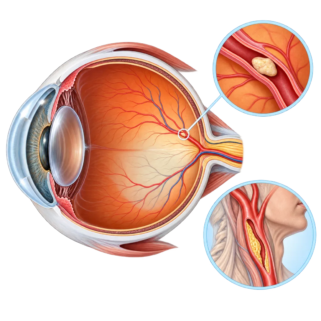
An eye stroke is a blockage of a retinal artery that causes sudden loss of vision in one eye, usually without pain. It is an emergency on par with a brain stroke: go to the emergency room right away.
Sources01 / Understanding the condition
About Eye Stroke
Eye stroke is the everyday name for a retinal artery occlusion. The retina is the light-sensitive layer at the back of the eyeball that turns light into signals for the brain. The artery that supplies the retina branches from the carotid artery, the vessel that also carries blood to the brain. For this reason, the 2021 AHA scientific statement regards retinal artery occlusion as a form of ischemic stroke. An overview of stroke is covered on the Stroke page.
Types of Blockage
When the blockage occurs in the central retinal artery, nearly all vision in one eye is lost within seconds. When a branch is blocked, only part of the visual field goes dark, for example the upper or lower half. Because it is painless, this symptom is often mistaken for tired eyes. There is also a temporary blockage, in which vision in one eye goes dark like a curtain coming down and then returns within a few minutes (amaurosis fugax). This is equivalent to a mini-stroke (transient ischemic attack) and is covered on the Minor Stroke and TIA page. Retinal vein occlusion, which is sometimes also called an eye stroke, is a different condition treated by an eye doctor.
Why You Should Go to the Emergency Room
Retinal cells begin to be damaged within a short time after blood flow stops, so the chance of saving vision depends on speed. In addition, the source of the blockage is often the same as the source of a brain stroke, and the risk of a brain stroke rises, especially in the first few weeks. For this reason, patients are advised to go directly to the emergency room so they can be assessed like stroke patients, rather than waiting for a regular eye appointment. In the emergency room, doctors also check for other signs of stroke in the face, arms, and speech.
Link to the Heart and Neck Arteries
The blockage is usually a fragment of plaque or a small clot carried by the bloodstream. Common sources are cholesterol plaque in the carotid arteries of the neck, covered on the Atherosclerosis page, and clots from the heart in atrial fibrillation or heart valve disease. Research shows that many patients only learn they have vascular risk factors, such as hypertension, diabetes, or carotid narrowing, after a full evaluation. In people over 50, doctors also consider an inflammation of the arteries called giant cell arteritis, because it requires prompt medicine to protect the other eye.
02 / Understanding the condition
Symptoms to Recognize.
Common Symptoms
Symptoms appear suddenly in one eye and are usually painless. Cover one eye at a time to find out which eye is affected.
- Sudden loss of vision or severely blurred vision in one eye
- No pain in the affected eye
- Part of the visual field in one eye goes dark, for example the upper or lower half
- Vision in one eye goes dark like a curtain coming down, then returns within a few minutes
- The eye looks normal from the outside even though vision is lost
Symptoms in Older Adults
In people over 50, the following symptoms may signal giant cell arteritis, an inflammation of the arteries that can also block the artery of the eye.
- A new headache at the temples
- A scalp that hurts when touched or when you comb your hair
- Jaw pain when chewing that improves when you stop
- Low-grade fever, weakness, and unexplained weight loss
- Pain and stiffness in the shoulders or hips in the morning
03 / Understanding the condition
What Causes This Condition?
- Fragments of plaque or clots from a carotid artery in the neck that has narrowed because of atherosclerosis
- Clots from the heart, for example in atrial fibrillation, heart valve disease, or after a heart attack
- Infected clumps from a heart valve, covered on the Infective Endocarditis page
- Inflammation of the artery walls in older age (giant cell arteritis)
- Blood clotting disorders, or clots that cross through a patent foramen ovale in younger people
- Rare complications of procedures, such as facial filler injections or blood vessel catheterization
04 / Understanding the condition
Who Is at Higher Risk?
Medical
Hypertension, diabetes, high LDL cholesterol, atrial fibrillation, heart valve disease, carotid artery narrowing, and a history of stroke or TIA. These are the same as the risk factors for a brain stroke.
Lifestyle
Smoking, lack of physical activity, a diet high in salt and saturated fat, and excess weight, which speed up plaque buildup in the blood vessels.
Age
It usually occurs after age 60. Giant cell arteritis almost always occurs after age 50.
Other
In younger people, rarer causes need to be looked for, such as blood clotting disorders, patent foramen ovale, autoimmune disease, or heart valve infection.
Groups at Higher Risk
- People with Carotid Artery Narrowing: Plaque in the neck arteries can release fragments into the arteries of the eye or brain, especially if it has already caused symptoms.
- People with Atrial Fibrillation or Valve Disease: Clots can form in the heart and then travel in the bloodstream to the retinal artery.
- Older Adults: The risk increases with age, and giant cell arteritis occurs almost only after age 50.
- People with Hypertension and Diabetes: Both damage the blood vessels and speed up plaque buildup in the carotid arteries.
05 / Understanding the condition
How Is It Diagnosed?
Sudden vision loss needs to be checked as quickly as possible, ideally in the emergency room of a hospital that can treat stroke. An eye doctor examines the retina to confirm an artery blockage: the retina looks pale and swollen, sometimes with a red spot in the center, and small fragments are sometimes visible inside the vessels. In people over 50, blood tests for inflammation are done the same day because giant cell arteritis requires prompt medicine.
Once the blockage is confirmed, the evaluation follows the approach for ischemic stroke: brain MRI, carotid artery testing, an ECG and heart rhythm recording, echocardiography, and blood tests for sugar and cholesterol. These tests should be completed within the first few days so the source of the blockage can be treated before a brain stroke occurs.
An eye examination by an ophthalmologist, including viewing the retina with an ophthalmoscope and OCT, to confirm an artery blockage and tell it apart from a vein blockage.
Brain MRI to look for small strokes that occurred at the same time without symptoms, which are found in some eye stroke patients.
Vascular Ultrasound
Ultrasound of the neck blood vessels, or CT angiography, to look for plaque and narrowing in the carotid arteries as the source of fragments.
Long-Term Heart Rhythm Monitoring
An ECG followed by rhythm recording for several days to several weeks to look for atrial fibrillation that comes and goes.
Echocardiography
Echocardiography to look for clots in the heart, valve abnormalities, valve infection, or a patent foramen ovale.
Erythrocyte Sedimentation Rate (ESR)
Erythrocyte sedimentation rate and CRP right away in people over 50 to look for signs of inflammation from giant cell arteritis.
Laboratory Tests
Blood tests for blood sugar, HbA1c, cholesterol, and clotting to assess vascular risk factors.
06 / Understanding the condition
Treatment Options.
Treatment is decided by a doctor after an assessment. The categories below can help you follow the conversation with your doctor.
Emergency Care as for a Stroke
You are assessed in the emergency room like a stroke patient. Tests to find the source of the blockage start right away, and preventive medicines are given according to the cause.
Clot-Dissolving Medicine (Thrombolysis)
For selected patients who arrive within the first few hours, doctors may consider a clot-dissolving medicine given through an IV. Its benefit is still being studied, so the decision is made together with the stroke team.
Steroids for Giant Cell Arteritis
If arteritis is suspected, steroid medicine is started right away, even before biopsy results are available, to protect the other eye. The type and duration are determined by your doctor.
Monitoring and Laser Treatment by an Eye Doctor
Regular follow-up visits to detect abnormal new vessels. If they appear, an eye doctor may use laser treatment on the retina or injections of medicine into the eye.
Preventing Another Stroke
Antiplatelets or anticoagulants depending on the cause, statins, blood pressure medicines, and carotid surgery for significant narrowing. The type of medicine is determined by your doctor.
07 / Understanding the condition
Complications to Watch For.
Permanent Vision Loss
Vision often does not fully recover, especially with a central artery blockage. An eye doctor can help you adjust with visual aids and training.
Brain Stroke
The risk of a brain stroke rises, especially in the first few weeks. Finding the source of the blockage and taking preventive medicines lower this risk.
Abnormal New Blood Vessels in the Eye
Weeks to months after the blockage, fragile vessels can grow and raise the pressure in the eye (neovascular glaucoma), so follow-up visits with an eye doctor need to be scheduled.
Heart Attack
People who have had an eye stroke often have blood vessel disease elsewhere, including the coronary arteries, so heart risk factors need to be controlled.
Damage to the Other Eye in Arteritis
If the cause is giant cell arteritis and it is not treated promptly, the other eye can also be affected within days.
08 / Understanding the condition
What Can You Do?
- Control your blood pressure, blood sugar, and cholesterol according to the targets set by your doctor
- Quit smoking and avoid secondhand smoke
- If you have atrial fibrillation, discuss blood thinners with your cardiologist
- Stay physically active for at least 150 minutes per week
- Get checked right away if vision in one eye goes dark briefly, even if it has returned
- Learn the stroke warning signs (BE FAST) and teach them to your family
09 / Understanding the condition
When Should You See a Doctor?
Sudden loss of vision in one eye is an emergency. Call 119 or go to an emergency room near you right away, even if your vision returns on its own within a few minutes, and note the time it started. Do not wait for a scheduled eye appointment or try eye drops on your own.
Make a consultation appointment if:
- You have had a brief episode of darkened vision in one eye that has not been checked
- You have just been discharged after an eye stroke and need to find the source of the blockage
- You often have palpitations with an irregular rhythm
- Your blood pressure, blood sugar, or cholesterol is not yet under control
10 / Common questions
Questions You May Have.
Frequently asked questions about eye stroke, its emergency signs, and its link to the heart.
Why is an eye stroke called a stroke?
The retinal artery is a branch of the blood vessel that also carries blood to the brain, and its blockage usually comes from the same sources as a brain stroke: plaque in the carotid artery or a clot from the heart. For this reason, retinal artery occlusion is regarded as a form of ischemic stroke. You need to be assessed like a stroke patient, including tests of the brain, neck vessels, and heart.
Can vision come back?
The chances depend on where the blockage is and how quickly blood flow returns. A branch artery blockage often leaves fairly good vision, while a central artery blockage less often recovers fully. Sometimes the blockage opens on its own within a few hours. Old methods such as massaging the eyeball have not been shown to help. What matters is going to the emergency room right away and preventing a brain stroke.
What is the difference between an eye stroke and retinal vein occlusion?
An artery blockage stops blood from flowing into the retina, so vision is lost suddenly and severely. A vein blockage slows the blood flowing out, so the retina swells and bleeds, with blurred vision that develops more gradually. Both are linked to hypertension and diabetes. Vein blockages are treated by an eye doctor, while artery blockages need emergency evaluation like a stroke.
My vision went dark briefly and then came back. Do I still need to go to the emergency room?
Yes. Vision in one eye that goes dark for a few minutes and then returns (amaurosis fugax) is equivalent to a mini-stroke (transient ischemic attack). It often signals plaque in the carotid artery or a clot from the heart, and a more serious stroke can follow within days. Prompt testing allows your doctor to find the source and give preventive medicine.
Why does a cardiologist take part in the evaluation after an eye stroke?
Some retinal artery blockages come from clots in the heart, especially in atrial fibrillation, valve disease, or valve infection. An irregular rhythm often comes and goes without symptoms, so it needs to be recorded over several days. Echocardiography looks for clots and valve problems. The results determine the choice of medicine: anticoagulants if the source is the heart, antiplatelets if the source is plaque.
What is giant cell arteritis?
Giant cell arteritis is inflammation of the walls of medium and large arteries, especially in the head, that almost always occurs after age 50. Symptoms include a new headache at the temples, jaw pain when chewing, and weakness. This inflammation can block the arteries of the eye. Steroid medicine needs to be started right away so that the other eye is not affected as well.
Sources and Medical Review
Translated from the Indonesian edition medically reviewed by dr. Indah A. Putri, Sp.N, Subsp, NIOO(K), MSc.
Sources as cited in the Indonesian edition.
- Mac Grory B, Schrag M, Biousse V, et al. Management of Central Retinal Artery Occlusion: A Scientific Statement From the American Heart Association. Stroke. 2021;52(6):e282-e294.
- Biousse V, Nahab F, Newman NJ. Management of Acute Retinal Ischemia: Follow the Guidelines! Ophthalmology. 2018;125(10):1597-1607.
- Schrag M, Youn T, Schindler J, Kirshner H, Greer D. Intravenous Fibrinolytic Therapy in Central Retinal Artery Occlusion: A Patient-Level Meta-analysis. JAMA Neurol. 2015;72(10):1148-54.
- Callizo J, Feltgen N, Pantenburg S, et al. Cardiovascular Risk Factors in Central Retinal Artery Occlusion: Results of a Prospective and Standardized Medical Examination. Ophthalmology. 2015;122(9):1881-8.
- Chodnicki KD, Pulido JS, Hodge DO, Klaas JP, Chen JJ. Stroke Risk Before and After Central Retinal Artery Occlusion in a US Cohort. Mayo Clin Proc. 2019;94(2):236-41.
- Kleindorfer DO, Towfighi A, Chaturvedi S, et al. 2021 Guideline for the Prevention of Stroke in Patients With Stroke and Transient Ischemic Attack: A Guideline From the American Heart Association/American Stroke Association. Stroke. 2021;52(7):e364-e467.
- Naylor R, Rantner B, Ancetti S, et al. European Society for Vascular Surgery (ESVS) 2023 Clinical Practice Guidelines on the Management of Atherosclerotic Carotid and Vertebral Artery Disease. Eur J Vasc Endovasc Surg. 2023;65(1):7-111.
Doctors Who Treat This Condition

dr. Indah A. Putri, Sp.N, Subsp, NIOO(K), MSc
Neurovascular, Interventional, Otology, Imaging, and Ophthalmology
Clinical focusStroke and Cerebrovascular Disease · Neurosonology
View Profile
dr. Emanoel Oepangat, Sp.JP(K)
Interventional Cardiology
Vascular Diagnostic and Therapeutic Center +1 center
Clinical focusCoronary Intervention · Peripheral Vascular Intervention
View Profile
dr. Sri Diniharini, Sp.JP(K)
Echocardiography & Cardiovascular Imaging
Cardiovascular Diagnostic Center +1 center
Clinical focusAdvanced Echocardiography · Transesophageal Echocardiography
View Profile