
Quick answer
What Is Paravalvular Leak?
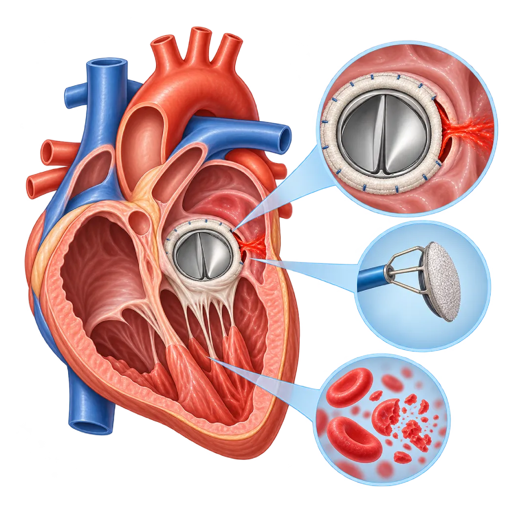
Paravalvular leak is a backflow of blood through a gap between the ring of an artificial heart valve and the surrounding heart tissue. Small leaks often cause no symptoms, while large ones can cause shortness of breath, anemia, and heart failure.
SourcesSigns That Need Urgent Help
- Severe shortness of breath at rest or waking up short of breath
- Coughing up pink, frothy sputum
- Chest pain, fainting, or cold and pale skin
- High fever and chills after a valve procedure
- Severe weakness with very dark urine
If these signs appear, go to the nearest emergency department right away or call Heart Attack Center 1500 258.
01 / Understanding the condition
About Paravalvular Leak
An artificial heart valve is sewn or implanted onto the ring of tissue where the original valve sat. If the valve does not seal tightly all the way around, a small gap forms between the ring of the artificial valve and the heart tissue. Blood can flow backward through this gap, outside the valve leaflets. This is called a paravalvular leak. Unlike a leak through the valve leaflets, this leak occurs at the edge of the valve. It can occur with both mechanical and biological valves, as a single gap or several gaps at once.
When a Leak Appears
A paravalvular leak can appear after valve replacement surgery, more often in the mitral position than in the aortic position, and after transcatheter aortic valve implantation (TAVI). In TAVI, the valve is not sewn in but pressed against the wall of the old valve, so uneven calcification can leave a gap. Some leaks are present from the time of the procedure, while others appear only years later, for example because stitches loosen or an infection damages the tissue around the valve.
Effects on the Heart and Blood
Doctors grade a leak as mild, moderate, or severe based on echocardiography. In most cases, the leak is mild and causes no symptoms. A moderate to severe leak makes the heart chamber take in extra blood with every beat, so over time the heart enlarges and weakens. Symptoms include shortness of breath, tiring easily, and swelling. In addition, blood that jets rapidly through a narrow gap can damage red blood cells (hemolysis). This leads to anemia with weakness, paleness, yellowing of the eyes, or dark urine. Hemolysis can occur even with a small gap, so the severity of symptoms does not always match the size of the gap.
Treatment Options
A mild leak without symptoms only needs monitoring with regular echocardiography. If the leak causes heart failure or hemolysis that requires transfusions, the gap can be closed with a small plug device through a catheter, covered on the Paravalvular Leak Closure page. Repeat surgery remains an option if there is an active infection, the valve is unstable, or the gap is not suitable for catheter closure. The decision is made by a heart team of imaging cardiologists, interventional cardiologists, and cardiac surgeons. Problems with artificial valves in general are covered on the Prosthetic Valve Dysfunction page.
02 / Understanding the condition
Symptoms to Recognize.
Common Symptoms
- Shortness of breath during activity or when lying down
- Tiring easily and a reduced ability to be active
- Leg swelling or rapid weight gain
- Palpitations or an irregular heart rhythm
- Weakness and paleness due to anemia
- Dark, tea-colored urine
- Yellowish eyes or skin
- A new murmur found by the doctor at a checkup
03 / Understanding the condition
What Causes This Condition?
- Heavy calcification of the valve ring that prevents the artificial valve from sealing tightly
- Fragile valve ring tissue, for example after repeated surgery
- Stitches that loosen or come undone over time
- Valve infection (endocarditis) that damages the tissue around the ring
- In TAVI: a valve size or position that does not fit the anatomy of the old valve well
04 / Understanding the condition
Who Is at Higher Risk?
Medical
Severe calcification of the valve ring, a history of endocarditis, repeated valve surgery, and connective tissue disease increase the chance of a gap around the valve.
Other
Valves in the mitral position develop paravalvular leaks more often than valves in the aortic position. In TAVI, uneven calcification and a poorly fitting valve size play a major role.
Age
Older adults more often have severe calcification and fragile tissue, and more often undergo TAVI.
Lifestyle
Poor dental hygiene increases the risk of endocarditis, one of the causes of leaks that appear later.
Groups at Higher Risk
- Patients After Mitral Valve Replacement: Paravalvular leaks are found more often in the mitral position, so follow-up echocardiography is important.
- Patients After TAVI: A mild leak after TAVI is fairly common; a moderate or larger leak needs further evaluation.
- Patients with a History of Endocarditis: Infection can damage the tissue around the valve ring and cause a new leak.
- Patients with Repeated Valve Surgery: Tissue that has been operated on several times tends to be more fragile for holding stitches.
05 / Understanding the condition
How Is It Diagnosed?
A paravalvular leak is often first suspected from a new murmur at a checkup, worsening shortness of breath, or unexplained anemia in a person with an artificial valve. Transthoracic echocardiography (TTE) is the first test to assess heart function and the direction of the leaking flow. Because the shadow of the artificial valve can obscure the image, transesophageal echocardiography (TEE), especially three-dimensional TEE, is used to see the location, number, shape, and size of the gaps.
Cardiac CT helps plan catheter closure, while cardiac MRI can measure the volume of leaking blood. Blood tests assess hemoglobin and markers of hemolysis such as LDH, haptoglobin, and bilirubin. If there is a fever, blood cultures are taken to rule out endocarditis before any procedure. The results of all tests are discussed by the heart team to choose between monitoring, catheter closure, or surgery.
Physical Examination
The doctor looks for a new murmur, signs of fluid buildup, and signs of anemia such as paleness or yellowing of the eyes.
Echocardiography
TTE assesses heart function and the severity of the leak; three-dimensional TEE maps the location, number, and size of the gaps.
Cardiac CT Scan
Maps the anatomy of the gap and the surrounding structures to plan catheter closure.
Cardiac MRI
Measures the volume of leaking blood and its effect on the size and function of the ventricles, especially for the aortic valve.
Complete Blood Count (CBC)
Assesses hemoglobin and reticulocytes to look for anemia caused by red blood cell damage.
Hemolysis markers (LDH, haptoglobin, and bilirubin) help confirm hemolysis and monitor its improvement after treatment.
Cardiac Catheterization
Measures the pressures in the heart chambers and assesses the coronary arteries when needed before a procedure.
06 / Understanding the condition
Treatment Options.
Treatment is decided by a doctor after an assessment. The categories below can help you follow the conversation with your doctor.
Regular Monitoring
A mild leak without symptoms and without hemolysis only needs monitoring with echocardiography on the schedule set by your doctor.
Heart Failure Medicines
Diuretics and other heart medicines to reduce shortness of breath and swelling; they do not close the gap, but they ease the load on the heart.
Anemia Treatment
Iron and folic acid supplements, plus transfusions if needed, depending on the severity of hemolysis and the doctor's decision.
Transcatheter Closure
A small plug device is inserted through a blood vessel or the heart wall to close the gap, guided by TEE; an option for selected patients with suitable anatomy.
Repeat Valve Surgery
Chosen when there is an active infection, an unstable valve, a very large gap or many gaps, or when transcatheter closure is not suitable.
Antibiotics for Endocarditis
If the leak is caused by infection, intravenous antibiotics are given for several weeks, and surgery is often needed.
07 / Understanding the condition
Complications to Watch For.
Heart Failure
The extra blood load makes the heart chambers enlarge and weaken, causing shortness of breath, swelling, and repeated hospital stays.
Hemolytic Anemia
Red blood cells are damaged as they pass through the narrow gap, causing weakness, paleness, dark urine, and sometimes a need for transfusions.
Pulmonary Hypertension
A long-standing mitral leak raises the pressure in the lung blood vessels and strains the right ventricle.
Heart Rhythm Disorders
Stretched upper chambers (atria) can trigger atrial fibrillation and an irregular heartbeat.
Reduced Kidney Function
Severe hemolysis and reduced blood flow can lower kidney function.
08 / Understanding the condition
What Can You Do?
- Have a baseline echocardiogram after valve replacement and attend regular checkups
- Keep your teeth clean and see a dentist regularly
- Tell your doctor that you have an artificial valve before any procedure
- Report any new shortness of breath, fatigue, or dark urine
- Have an unexplained fever checked before taking antibiotics
- Take your medicines as advised and attend checkups on time
09 / Understanding the condition
When Should You See a Doctor?
If you have an artificial heart valve, whether placed through surgery or TAVI, make an earlier appointment if your shortness of breath and fatigue increase, your legs start to swell, or you look pale or have yellowish eyes or dark, tea-colored urine. Also report it if lab results show a drop in hemoglobin or if you start to need transfusions more often.
Go to the emergency room right away if you have:
- Severe shortness of breath at rest or waking up short of breath
- A cough with pink, frothy sputum
- Chest pain, fainting, or cold and pale skin
- High fever and chills after a valve procedure
Don't Wait in an Emergency.
If symptoms are severe or come on suddenly, go to the nearest emergency department right away or call Heart Attack Center 1500 258, rather than waiting for an appointment.
10 / Common questions
Questions You May Have.
Frequently asked questions about leaks around an artificial heart valve.
Do all paravalvular leaks need to be closed?
No. Many leaks are mild, cause no symptoms, and do not damage red blood cells. Leaks like these only need monitoring with regular echocardiography. Closure is considered when the leak causes heart failure, hemolysis that requires transfusions, or growing enlargement of the heart. The decision is made by the heart team based on the size and location of the gap and your overall condition.
What is the difference between catheter closure and repeat surgery?
Catheter closure places a small plug device through a blood vessel or the heart wall without opening the chest, so recovery is usually shorter. This approach is suitable if the gap can be reached and there is no active infection. Repeat surgery lets the doctor re-stitch or replace the valve directly, and it is chosen if the valve is unstable, there is an infection, or the gap is too large.
Why can a small leak make me anemic?
Blood pushed by the heart jets very fast through a narrow gap. This friction and pressure can tear red blood cells, which is called hemolysis. Because the jet is stronger, a small gap sometimes causes more noticeable hemolysis than a large gap. Signs include weakness, paleness, yellowing of the eyes, and dark urine. When severe, hemolysis is a strong reason to close the gap.
Is a leak after TAVI the same as a leak after surgery?
The principle is the same: blood leaks through a gap at the edge of the artificial valve. In TAVI, the valve is not sewn in but pressed against the old valve, so uneven calcification can leave a gap. A mild leak after TAVI is fairly common and is usually only monitored. A moderate or severe leak is linked to less favorable long-term outcomes, so it needs evaluation by the heart team.
Does catheter closure always eliminate the leak?
Not always completely. The goal is to reduce the leak to a level that no longer strains the heart or damages red blood cells. Gaps with an irregular shape can sometimes be closed only partly, and some people need an additional procedure. After the procedure, echocardiography and blood tests are repeated to assess any remaining leak and the improvement in hemolysis.
Sources and Medical Review
Translated from the Indonesian edition medically reviewed by Dr. dr. Dafsah A. Juzar, Sp.JP(K).
Sources as cited in the Indonesian edition.
- Ruiz CE, Hahn RT, Berrebi A, et al. Clinical Trial Principles and Endpoint Definitions for Paravalvular Leaks in Surgical Prosthesis: An Expert Statement. J Am Coll Cardiol. 2017;69(16):2067-2087.
- Zoghbi WA, Jone PN, Chamsi-Pasha MA, et al. Guidelines for the Evaluation of Prosthetic Valve Function With Cardiovascular Imaging: A Report From the American Society of Echocardiography Developed in Collaboration With the Society for Cardiovascular Magnetic Resonance and the Society of Cardiovascular Computed Tomography. J Am Soc Echocardiogr. 2024;37(1):2-63.
- Praz F, Borger MA, Lanz J, et al. 2025 ESC/EACTS Guidelines for the management of valvular heart disease. Eur Heart J. 2025;46(44):4635-4736.
- Otto CM, Nishimura RA, Bonow RO, et al. 2020 ACC/AHA Guideline for the Management of Patients With Valvular Heart Disease: A Report of the American College of Cardiology/American Heart Association Joint Committee on Clinical Practice Guidelines. Circulation. 2021;143(5):e72-e227.
- Genereux P, Piazza N, Alu MC, et al. Valve Academic Research Consortium 3: updated endpoint definitions for aortic valve clinical research. Eur Heart J. 2021;42(19):1825-1857.
- Millan X, Skaf S, Joseph L, et al. Transcatheter Reduction of Paravalvular Leaks: A Systematic Review and Meta-analysis. Can J Cardiol. 2015;31(3):260-269.
Doctors Who Treat This Condition

dr. BRM Ario S. Kuncoro, Sp.JP(K)
Echocardiography & Cardiovascular Imaging
Cardiovascular Diagnostic Center +2 centers
Clinical focusAdvanced and 3D Echocardiography · Transesophageal Echocardiography
View Profile
Dr. dr. Faris Basalamah, Sp.JP(K)
Interventional Cardiology
Coronary Intervention Procedures +1 center
Clinical focusComplex Coronary Intervention · Cardiac Electrophysiology
View Profile
Dr. dr. Dafsah A. Juzar, Sp.JP(K)
Interventional Cardiology
Coronary Intervention Procedures +1 center
Clinical focusComplex Coronary Intervention · Cardiac Catheterization
View Profile