
Quick answer
What Is Paravalvular Leak Closure?
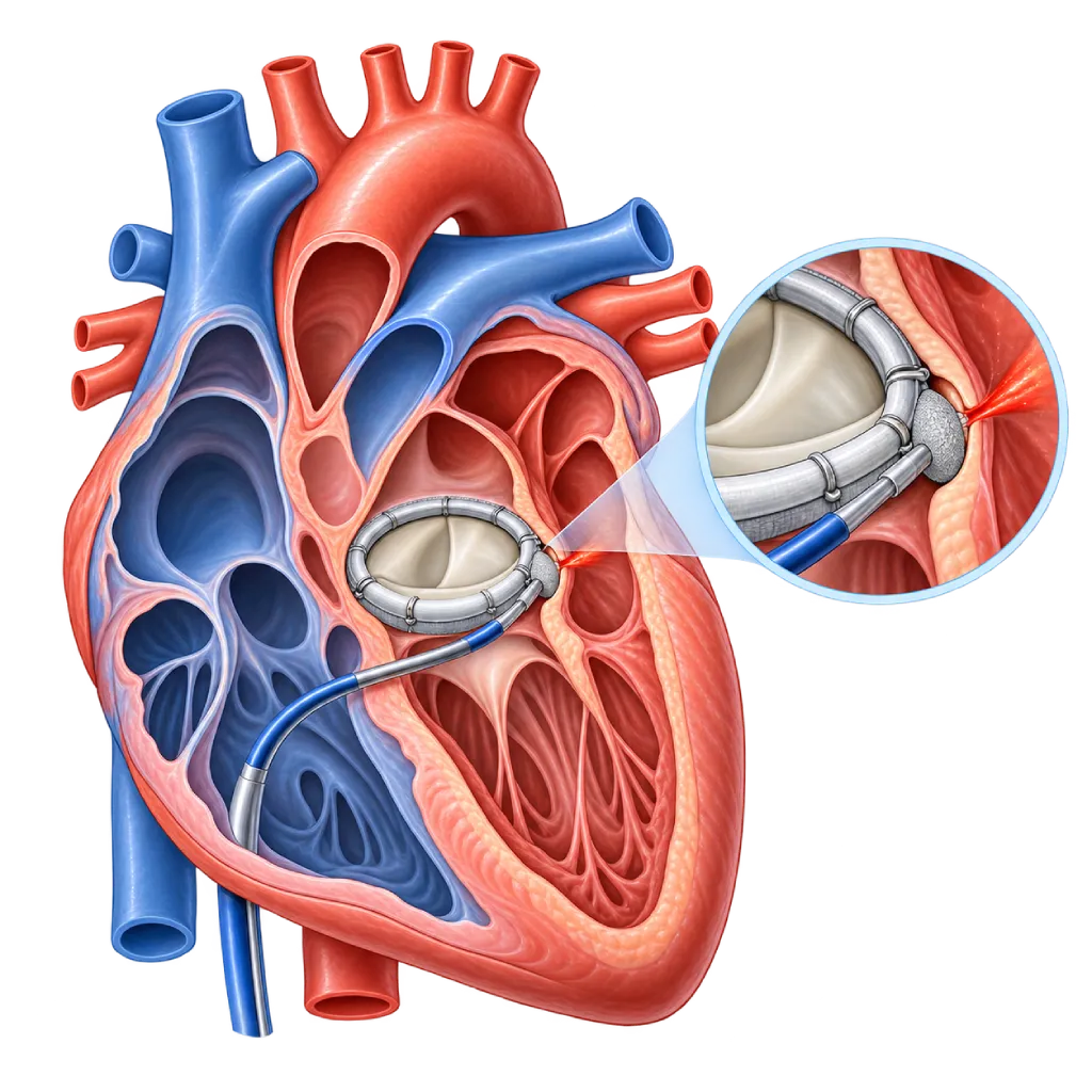
Paravalvular leak closure is a procedure to close a gap between an artificial heart valve and the surrounding heart tissue, which lets blood leak beside the valve. The doctor places a small plugging device through a catheter, guided by transesophageal echocardiography. The procedure usually takes 1 to 3 hours, sometimes longer.
Sources01 / Understanding the procedure
About Paravalvular Leak Closure
What Is Paravalvular Leak Closure
An artificial heart valve is sewn or expanded onto the original valve ring. Sometimes a gap forms between the edge of the artificial valve and the surrounding heart tissue, for example because of loose stitches, fragile tissue, calcification, or infection. Blood then leaks through that gap, outside the valve leaflets. This condition is called a paravalvular leak, or a leak around the valve.
Paravalvular leak closure is a procedure to plug that gap with a small device inserted through a catheter, as an alternative to repeat surgery.
When a Leak Needs to Be Closed
Small leaks often cause no symptoms and only need monitoring with echocardiography. Larger leaks can cause heart failure, with shortness of breath, tiring easily, and swelling. Blood jetting through a narrow gap can also damage red blood cells (hemolysis), which can cause anemia, dark tea-colored urine, and the need for repeated transfusions.
How It Works in Brief
The catheter is guided to the gap using transesophageal echocardiography (TEE) and X-rays. After a guidewire crosses the gap, a plugging device in the form of a plug or disc made of woven metal mesh (occluder), or sometimes a small wire coil, is expanded to close it. One gap may need more than one device, and several gaps can be closed in one procedure.
The Catheter Route to the Valve
For the aortic valve, the catheter is usually inserted through an artery in the groin. For the mitral valve, the catheter is inserted through a vein in the groin and then passed through the wall between the right and left atria. In a small number of cases, the doctor uses another route, for example a small puncture in the chest wall toward the tip of the heart.
Catheter Closure or Repeat Surgery
Repeat surgery to stitch the gap closed or replace the valve, covered on the Heart Valve Surgery page, remains an option, especially when the gap is very large, the artificial valve is unstable, or there is an infection. Catheter closure is considered when the risk of repeat surgery is high and the shape of the gap is suitable. The decision is made by the heart team together with you.
A leak around the valve can also occur after the procedure covered on the Transcatheter Aortic Valve Implantation (TAVI) page. In some of these patients, the gap can be closed in a similar way.
02 / Understanding the procedure
When Is This Procedure Considered?
A doctor may consider this procedure in the situations below. The decision is always made after a thorough assessment.
- Heart Failure Symptoms Due to the LeakShortness of breath, tiring easily, or swelling related to a moderate to severe leak around the valve.
- Anemia from Red Blood Cell DamageHemolysis that causes severe anemia or a need for repeated transfusions.
- High Risk of Repeat SurgeryA history of several heart operations, older age, or other illnesses that make surgery risky.
- Suitable Gap ShapeThe gap is not too large and the artificial valve remains firmly in place.
- Leak Around a TAVI ValveIn some patients, a significant leak after TAVI can be closed with a similar device.
Situations to Consider First
- Active Valve InfectionEndocarditis needs to be treated with antibiotics and often surgery, not with a plugging device.
- Unstable Artificial ValveIf the artificial valve moves abnormally because the gap is very large, repeat surgery is more suitable.
- Clot in the Heart or on the ValveA clot needs to be treated first because it can break loose when the catheter passes it.
- Gap That Could Interfere with the ValveWhen a device could block the movement of the artificial valve leaflets or cannot close the gap well.
03 / Before the procedure
How Should You Prepare?
Paravalvular leak closure is usually a planned procedure after evaluation by the heart team.
Transesophageal echocardiography. A small probe is passed through the mouth into the esophagus to see the location, size, and number of gaps. Three-dimensional images help the team understand the shape of the gap.
Cardiac CT scan. In some patients, CT helps measure the gap and plan the catheter route.
Blood tests. A complete blood count, markers of red blood cell damage, kidney function, and INR for people taking warfarin.
Blood thinners. People with a mechanical valve usually take warfarin. The dose is adjusted before the procedure according to the team's instructions. Do not stop or change the dose on your own.
Fasting and consent. Follow the fasting instructions before general anesthesia. Before you sign the consent form, your doctor explains the benefits, the risks, the possibility that the gap cannot be closed completely, and the option of repeat surgery.
Unsure about the preparation instructions? Ask our team before the procedure.
04 / During the procedure
How Is the Procedure Done?
The procedure is performed in the cardiac catheterization lab by an interventional cardiologist together with an echocardiography specialist, usually under general anesthesia so the TEE probe can be used throughout the procedure. It takes about 1 to 3 hours and can take longer if the gap is complex.
Locating the Gap
TEE and X-rays show where the gap is in relation to the artificial valve, so the doctor can choose the direction and route of the catheter.
Crossing the Gap
The doctor guides a catheter and a very thin guidewire across the gap, through an artery or vein in the groin, and then a delivery catheter is placed inside the gap.
Placing the Plugging Device
The plugging device is delivered through the catheter and expanded inside the gap. Before the device is released, the team makes sure it does not interfere with the movement of the artificial valve leaflets.
Checking the Result and Closing the Puncture
Any remaining leak is assessed with TEE. If needed, a second device is placed. The catheter is then removed and the puncture in the groin is closed.
05 / Benefits and risks
Benefits and Risks Should Be Discussed Together.
Every medical procedure has benefits and risks. Your doctor will explain both for your situation before the procedure.
Benefits You Can Expect
- No Repeat Heart SurgeryThe gap is closed through a blood vessel, so the chest does not need to be opened again.
- Heart Failure Symptoms Can EaseIn many patients, shortness of breath and tiring easily improve after the leak becomes smaller.
- Anemia Can ImproveThe need for transfusions can decrease as red blood cell damage subsides.
- Shorter RecoveryUsually a hospital stay of a few days, shorter than after repeat surgery.
The benefits of a procedure are not a promise of a cure. Ask how the results will affect your care plan.
Risks You Should Know
- Remaining LeakThe gap cannot always be closed completely; some patients need an additional device or surgery.
- Device Interfering with the Artificial ValveThe device can block the movement of the valve leaflets; that is why its position is checked with TEE before it is released.
- Device DisplacementRarely, the device can come out of the gap and need to be retrieved through a catheter or surgery.
- Increased HemolysisA small remaining leak around the device can temporarily worsen red blood cell damage.
- StrokeClots or air can travel to the brain during the procedure; the team takes preventive measures.
- Bleeding and Puncture Site ProblemsMore likely in people taking blood thinners, for example bruising, bleeding, or a tear in the blood vessel.
Risks can differ from person to person. A full explanation and consent are given after an individual assessment.
06 / After the procedure
What Is Recovery Like?
Patients usually stay in the hospital for 1 to 3 days, depending on their condition before the procedure.
Monitoring. Heart rhythm, the puncture site, and signs of red blood cell damage are monitored. In some patients, hemolysis can temporarily increase if a small leak remains around the device.
Medications. Blood thinners for a mechanical valve are continued as advised. Heart failure medications and iron supplements are given if needed.
Follow-up visits. Echocardiography and blood tests are done about one month after the procedure, then periodically, to assess any remaining leak and improvement in anemia.
Activity. Light activities can be started within a few days. Avoid lifting heavy objects for about one week or as advised by your doctor.
Warning signs. Go to the emergency room right away if you have:
- Shortness of breath or leg swelling that gets worse
- Dark tea-colored urine, yellowish eyes, or feeling very weak and pale
- Fever with no clear cause
- Facial drooping, weakness on one side of the body, or slurred speech
- Bleeding or swelling that grows larger in the groin
- A mechanical valve click that changes or cannot be heard as usual
Know the Signs That Need Prompt Care.
Follow the team's guidance on warning signs. If severe or sudden symptoms appear after the procedure, go to the nearest emergency department right away or call Heart Attack Center 1500 258.
07 / Team and support
Doctors and Procedure Support
The doctors below are listed for this procedure. Listed by name, not by ranking.

Dr. dr. Dafsah A. Juzar, Sp.JP(K)
Interventional Cardiology
Coronary Intervention Procedures +1 center
Clinical focusComplex Coronary Intervention · Cardiac Catheterization
View Profile
Dr. dr. Faris Basalamah, Sp.JP(K)
Interventional Cardiology
Coronary Intervention Procedures +1 center
Clinical focusComplex Coronary Intervention · Cardiac Electrophysiology
View Profile
dr. Sri Diniharini, Sp.JP(K)
Echocardiography & Cardiovascular Imaging
Cardiovascular Diagnostic Center +1 center
Clinical focusAdvanced Echocardiography · Transesophageal Echocardiography
View ProfileCatheterization Laboratory (Cath Lab)
A dedicated procedure room for cardiac catheterization, coronary angiography, stent placement, and other catheter-based heart…
4D Echocardiography
An ultrasound scan of the heart that records three-dimensional images in real time to assess pumping function, the valves, and…
The availability of technology and test support is confirmed with the team. Technology is chosen according to clinical need.
08 / Common questions
Before You Plan the Procedure.
Questions people with artificial heart valves often ask about paravalvular leak closure.
How do I know if there is a leak around my artificial valve?
Small leaks often cause no symptoms and are only seen on a follow-up echocardiogram. Larger leaks can cause shortness of breath, tiring easily, leg swelling, or anemia with dark urine. Your doctor may also hear a new heart murmur. Transesophageal echocardiography confirms the location and size of the gap. That is why regular follow-up after valve surgery is important.
Do all paravalvular leaks need to be closed?
No. Small leaks without symptoms and without anemia are usually just monitored periodically with echocardiography and blood tests. Closure is considered when the leak causes heart failure symptoms or damages red blood cells to the point of causing anemia. The heart team weighs the size of the gap, your symptoms, and the risks of the procedure before recommending closure or repeat surgery.
Is this procedure painful?
The procedure is usually done under general anesthesia, so you are asleep and do not feel anything. After waking up, your groin may feel sore and slightly bruised, and your throat may feel a little uncomfortable from the TEE probe for one to two days. These symptoms are usually relieved with ordinary pain medication.
How often is closure successful?
Studies show that the leak can be reduced in the majority of patients, and many of them experience improvement in heart failure symptoms or anemia. The results depend on the location, shape, and number of gaps, as well as the type of artificial valve. Some patients still have a remaining leak or need further treatment. Your doctor will explain what to expect in your situation.
How is it different from repeat surgery?
Repeat surgery reopens the chest to stitch the gap closed or replace the valve, so recovery takes longer and the risk is higher in patients who have had several previous operations. Catheter closure does not open the chest, but it is not suitable for every gap, for example when the valve is unstable or there is an infection. The surgery is covered on the Heart Valve Surgery page.
Do I still take warfarin after the procedure?
Yes, if you have a mechanical valve, warfarin is still taken for life because it is needed to prevent clots on the valve. The dose may be adjusted temporarily around the procedure, then returned to your target INR. People with a biological valve follow their doctor's medication advice. Do not stop or change the dose without consulting your doctor.
Sources and Medical Review
Translated from the Indonesian edition medically reviewed by dr. Sri Diniharini, Sp.JP(K).
Sources as cited in the Indonesian edition.
- Vahanian A, Beyersdorf F, Praz F, et al. 2021 ESC/EACTS Guidelines for the management of valvular heart disease. Eur Heart J. 2022;43(7):561-632.
- Otto CM, Nishimura RA, Bonow RO, et al. 2020 ACC/AHA Guideline for the Management of Patients With Valvular Heart Disease: A Report of the American College of Cardiology/American Heart Association Joint Committee on Clinical Practice Guidelines. Circulation. 2021;143(5):e72-e227.
- Ruiz CE, Hahn RT, Berrebi A, et al. Clinical trial principles and endpoint definitions for paravalvular leaks in surgical prosthesis: an expert statement. J Am Coll Cardiol. 2017;69(16):2067-2087.
- Zoghbi WA, Jone PN, Chamsi-Pasha MA, et al. Guidelines for the Evaluation of Prosthetic Valve Function With Cardiovascular Imaging: A Report From the American Society of Echocardiography. J Am Soc Echocardiogr. 2024;37(1):2-63.
- Sorajja P, Cabalka AK, Hagler DJ, Rihal CS. Percutaneous repair of paravalvular prosthetic regurgitation: acute and 30-day outcomes in 115 patients. Circ Cardiovasc Interv. 2011;4(4):314-321.
- Sorajja P, Cabalka AK, Hagler DJ, Rihal CS. Long-term follow-up of percutaneous repair of paravalvular prosthetic regurgitation. J Am Coll Cardiol. 2011;58(21):2218-2224.
- Millán X, Skaf S, Joseph L, et al. Transcatheter reduction of paravalvular leaks: a systematic review and meta-analysis. Can J Cardiol. 2015;31(3):260-269.