
Quick answer
What Is Constrictive Pericarditis?
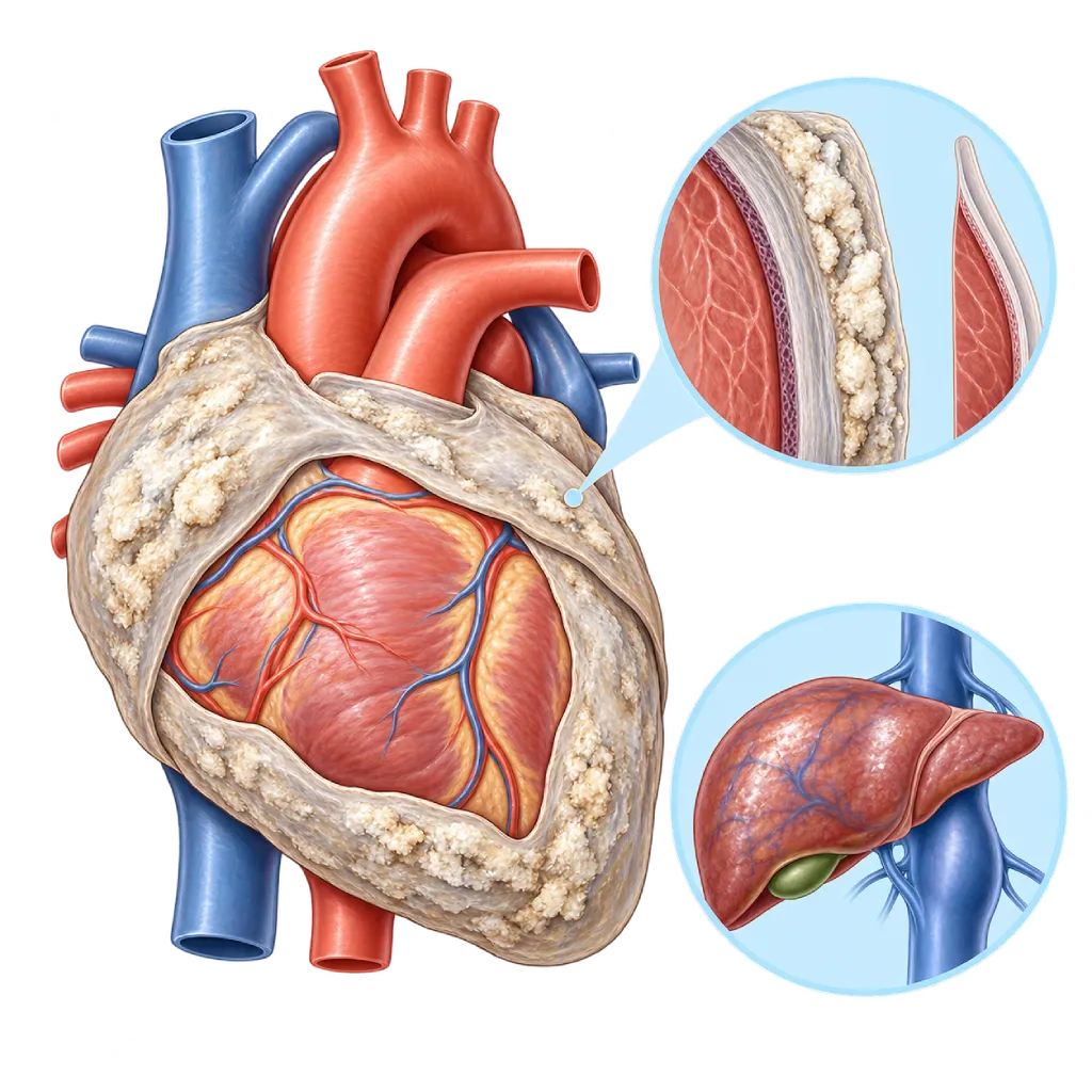
Constrictive pericarditis is a condition in which the sac around the heart becomes thick, stiff, and sometimes calcified, so the heart cannot fully expand. This causes swelling, an enlarged abdomen, and shortness of breath similar to heart failure.
SourcesSigns That Need Urgent Help
- Severe shortness of breath, including at rest or when lying down
- Fainting or nearly fainting
- Swelling and abdominal enlargement that increase quickly
- A very fast or irregular heartbeat with weakness
- Passing very little urine
- Confusion, severe weakness, or cold and pale skin
If these signs appear, go to the nearest emergency department right away or call Heart Attack Center 1500 258.
01 / Understanding the condition
About Constrictive Pericarditis
Normally the pericardium is flexible, so the heart can expand freely to take in blood between beats. In constrictive pericarditis, long-lasting or repeated inflammation turns the pericardium into thick, stiff scar tissue, sometimes containing calcium. This membrane wraps around the heart like a shell. The heart muscle itself is often still strong, but the heart chambers cannot fill completely.
Because filling is restricted, blood backs up in the veins returning to the heart. Fluid builds up in the legs, abdomen, liver, and the space around the lungs. The veins in the neck look swollen and sometimes bulge even more when you breathe in. Symptoms usually develop slowly over months, so they are often mistaken for liver or kidney disease.
Causes in Indonesia and Worldwide
In developed countries, causes include viral or idiopathic pericarditis, heart surgery, and radiation therapy to the chest. In countries with many tuberculosis (TB) cases, including Indonesia, TB is an important cause. The risk of constriction after TB pericarditis or a pus-forming bacterial infection is much higher than after viral pericarditis.
Three Forms of Constriction
Doctors distinguish transient constriction, effusive constriction, and chronic constriction. Transient constriction occurs while inflammation is still active and can improve with anti-inflammatory medicines within two to three months. Effusive constriction is a combination of fluid and a stiff membrane, so pressure in the heart stays high even after the fluid has been drained; fluid in the sac around the heart is covered on the Pericardial Effusion page. Chronic constriction means the membrane has become permanent scar tissue; the main treatment is surgery to remove the pericardium, called pericardiectomy.
Similar to Restrictive Cardiomyopathy
The symptoms of constrictive pericarditis are similar to those of a condition in which the heart muscle is stiff, covered on the Restrictive Cardiomyopathy page. Telling the two apart matters because constriction can be treated with surgery, while restrictive cardiomyopathy cannot. That is why doctors usually combine several tests before deciding on treatment.
Most patients who undergo pericardiectomy find that their symptoms ease. Results tend to be better if surgery is done before the heart and liver are damaged by long-term congestion. While a decision is being made together with the heart team and the surgical team, diuretics (water pills) help relieve swelling and shortness of breath.
02 / Understanding the condition
Symptoms to Recognize.
Common Symptoms
- Swelling in the legs and ankles
- An enlarged abdomen from fluid (ascites) and a bloated feeling
- Tiring quickly and feeling weak
- Shortness of breath during activity
- Swollen-looking veins in the neck
- A feeling of fullness or dull pain in the upper right abdomen from a swollen liver
- Loss of appetite and visible muscle wasting
- Palpitations or an irregular pulse
03 / Understanding the condition
What Causes This Condition?
- Pericarditis caused by tuberculosis (TB), an important cause in countries with many TB cases
- Viral or idiopathic pericarditis that leaves scar tissue
- Previous heart surgery
- Radiation therapy to the chest area, for example for breast cancer, lung cancer, or lymphoma
- A pus-forming bacterial infection of the heart's lining (purulent pericarditis)
- Autoimmune diseases such as lupus and rheumatoid arthritis
- Severe kidney failure, cancer in the pericardium, or a chest injury
04 / Understanding the condition
Who Is at Higher Risk?
Medical
A history of pericarditis, especially if caused by TB or a bacterial infection, previous heart surgery, radiation therapy to the chest, autoimmune disease, and kidney failure.
Other
Living in an area with many TB cases, close contact with people who have TB, incomplete TB treatment, and HIV or other conditions that weaken the immune system.
Groups at Higher Risk
- TB Pericarditis Survivors: The risk of constriction after TB pericarditis is much higher than after viral pericarditis, so monitoring is needed after treatment.
- Patients Who Have Had Heart Surgery: Scar tissue after surgery can make the pericardium stiff, sometimes years later.
- Cancer Survivors with Chest Radiation: Radiation can damage the pericardium and cause constriction years after treatment, often together with valve and heart muscle problems.
- Purulent Pericarditis Survivors: A pus-forming bacterial infection of the heart's lining leaves a high risk of constriction even after the infection has healed.
05 / Understanding the condition
How Is It Diagnosed?
Your doctor suspects constrictive pericarditis when there are signs of congestion, such as swelling, an enlarged abdomen, and swollen neck veins, even though the heart's pumping strength is still good. A history of pericarditis, TB, heart surgery, or chest radiation strengthens the suspicion.
Echocardiography is usually the first test, used to look for the typical signs of constriction. A cardiac CT scan assesses the thickness and calcification of the membrane, while a cardiac MRI can also show active inflammation. Knowing whether inflammation is active is important because this type of constriction can improve with medicine. If imaging results are still unclear, cardiac catheterization measures the pressures in the right and left heart chambers at the same time. In cases that are hard to tell apart from restrictive cardiomyopathy, doctors sometimes need a heart muscle biopsy. Tests for the cause, including TB, are done based on your history and findings.
Echocardiography
Looking for typical signs, such as a bouncing motion of the wall between the ventricles (septal bounce), changes in blood flow with breathing, and widened large veins.
Cardiac MRI
Measuring the thickness of the pericardium, showing active inflammation that may still improve with medicine, and assessing how the two ventricles affect each other during breathing.
Cardiac CT Scan
Showing thickening and calcification of the pericardium, useful for confirming the diagnosis and planning surgery.
Chest X-Ray
Sometimes shows a line of calcium around the heart and fluid in the pleural space around the lungs.
Cardiac Catheterization
Measuring pressures in the right and left heart chambers to confirm constriction and tell it apart from restrictive cardiomyopathy when imaging is unclear.
NT-proBNP and BNP Test
Levels tend to be lower in constriction than in restrictive cardiomyopathy, which helps tell the two apart.
C-Reactive Protein (CRP) Test
A high CRP shows that inflammation is still active, a clue that the constriction may be transient and can improve with medicine.
06 / Understanding the condition
Treatment Options.
Treatment is decided by a doctor after an assessment. The categories below can help you follow the conversation with your doctor.
Anti-Inflammatory Medicines for Transient Constriction
If inflammation is still active, anti-inflammatory medicines, colchicine, or corticosteroids can reverse the constriction within a few months, so surgery is not always needed.
Tuberculosis Treatment
Anti-TB drugs are given if the cause is TB, to treat the infection and lower the risk of permanent constriction. Your doctor may add other medicines depending on your condition.
Diuretics to Reduce Congestion
Diuretics help reduce swelling and shortness of breath, but they only relieve symptoms and do not remove the stiff membrane.
Pericardiectomy
Surgery to remove most of the stiff pericardium so the heart can expand again. This is the main treatment for chronic constriction and is performed by a heart surgery team.
Regular Monitoring
Patients who have not yet had surgery, or who cannot have it, are monitored with echocardiography and blood tests to assess changes in congestion and organ function.
07 / Understanding the condition
Complications to Watch For.
Chronic Heart Failure
Restricted filling of the heart causes ongoing fluid congestion, shortness of breath, and fatigue that tend to get worse if not treated.
Impaired Liver Function
Long-term congestion of blood in the liver can impair its function, lower albumin levels, and worsen swelling and ascites.
Atrial Fibrillation
Upper heart chambers that are enlarged by high pressure make an irregular rhythm more likely, covered on the Atrial Fibrillation page.
Intestinal Protein Loss
Congestion in the blood vessels of the intestines can make protein leak through the digestive tract, so muscles waste away and swelling increases.
Long-Term Heart Muscle Damage
Constriction that lasts for years can weaken the heart muscle, so surgery offers less benefit if treatment is delayed.
08 / Understanding the condition
What Can You Do?
- Complete your TB treatment fully, following your doctor's schedule
- Treat pericarditis completely and do not stop your medicines on your own
- Go for regular checkups if you have had TB pericarditis or pus-forming pericarditis
- If you have had chest radiation, mention this history and have your heart checked regularly as advised
- If you have been diagnosed, limit salt and weigh yourself every morning
- See a doctor if you have swelling and an enlarging abdomen without a clear cause
09 / Understanding the condition
When Should You See a Doctor?
See a doctor if you have leg swelling, an enlarging abdomen, tiring quickly, or shortness of breath that gets worse over several weeks to months, especially if you have had pericarditis, TB, heart surgery, or chest radiation. These symptoms are often mistaken for liver or kidney disease, so tell your doctor about this history. If you have already been diagnosed, regular checkups help determine the right time for treatment.
Go to the emergency room right away if you have:
- Severe shortness of breath, including at rest or when lying down
- Fainting or nearly fainting
- A very fast or irregular heartbeat with weakness
- Swelling and abdominal enlargement that increase quickly, with very little urine
Don't Wait in an Emergency.
If symptoms are severe or come on suddenly, go to the nearest emergency department right away or call Heart Attack Center 1500 258, rather than waiting for an appointment.
10 / Common questions
Questions You May Have.
Common questions about constrictive pericarditis.
Can constrictive pericarditis get better without surgery?
Some cases can. If tests show that inflammation is still active, for example a high CRP or an MRI showing an inflamed membrane, the constriction may be transient and improve with anti-inflammatory medicines within a few months. If the membrane has already become thick scar tissue or calcified, medicines only relieve symptoms, and surgery to remove the pericardium becomes the main option to restore the heart's filling.
What is pericardiectomy surgery like?
Pericardiectomy is heart surgery to remove most of the stiff pericardium, usually through an incision in the breastbone. Once the membrane is removed, the heart can expand and fill with blood again. Many patients notice less swelling and shortness of breath, although full recovery can take several weeks to months. The surgical team will explain the risks and preparation that suit your condition.
What is the difference between constrictive pericarditis and restrictive cardiomyopathy?
Both make it hard for the heart to fill with blood and cause similar symptoms. In constrictive pericarditis, the problem is a stiff outer membrane, while the heart muscle is usually still normal. In restrictive cardiomyopathy, the heart muscle itself is stiff. The difference matters because constriction can be treated with surgery. Echocardiography, MRI, a BNP test, and catheterization help tell them apart.
Why can TB cause constrictive pericarditis?
TB can spread to the pericardium and cause long-lasting inflammation and thick fluid. As it heals, this inflammation often leaves thick scar tissue that binds the heart. This risk can be reduced by finding and treating TB pericarditis as early as possible and completing anti-TB treatment on schedule. Monitoring after treatment helps detect constriction earlier.
Do I need to limit salt and fluids?
When constriction causes swelling, limiting salt helps reduce fluid buildup, and your doctor usually prescribes diuretics. Fluid limits depend on each person's condition, so ask about the right limit for you. Weighing yourself every morning helps detect fluid buildup early; rapid weight gain over a few days should be reported to your doctor.
Sources and Medical Review
Translated from the Indonesian edition medically reviewed by dr. Sri Diniharini, Sp.JP(K).
Sources as cited in the Indonesian edition.
- Schulz-Menger J, Collini V, Gröschel J, Adler Y, Brucato A, Christian V, et al. 2025 ESC Guidelines for the management of myocarditis and pericarditis. Eur Heart J. 2025;46(40):3952-4041.
- Adler Y, Charron P, Imazio M, Badano L, Barón-Esquivias G, Bogaert J, et al. 2015 ESC Guidelines for the diagnosis and management of pericardial diseases: The Task Force for the Diagnosis and Management of Pericardial Diseases of the European Society of Cardiology (ESC). Eur Heart J. 2015;36(42):2921-2964.
- Welch TD. Constrictive pericarditis: diagnosis, management and clinical outcomes. Heart. 2018;104(9):725-731.
- Syed FF, Schaff HV, Oh JK. Constrictive pericarditis: a curable diastolic heart failure. Nat Rev Cardiol. 2014;11(9):530-544.
- Welch TD, Ling LH, Espinosa RE, Anavekar NS, Wiste HJ, Lahr BD, et al. Echocardiographic Diagnosis of Constrictive Pericarditis: Mayo Clinic Criteria. Circ Cardiovasc Imaging. 2014;7(3):526-534.
- Ling LH, Oh JK, Schaff HV, Danielson GK, Mahoney DW, Seward JB, et al. Constrictive Pericarditis in the Modern Era: Evolving Clinical Spectrum and Impact on Outcome After Pericardiectomy. Circulation. 1999;100(13):1380-1386.
- Haley JH, Tajik AJ, Danielson GK, Schaff HV, Mulvagh SL, Oh JK. Transient constrictive pericarditis: causes and natural history. J Am Coll Cardiol. 2004;43(2):271-275.
Doctors Who Treat This Condition

dr. Sri Diniharini, Sp.JP(K)
Echocardiography & Cardiovascular Imaging
Cardiovascular Diagnostic Center +1 center
Clinical focusAdvanced Echocardiography · Transesophageal Echocardiography
View Profile
Dr. dr. Dudy A. Hanafy, Sp.BTKV, Subsp. JD(K), MARS
Adult Cardiac Surgery
Advanced Cardiovascular Surgical Care +1 center
Clinical focusMinimally Invasive Cardiac Surgery · Coronary Artery Bypass Surgery
View Profile
dr. Radityo Prakoso, Sp.JP(K)
Pediatrics & Congenital Heart Disease
Congenital and Structural Heart Center
Clinical focusPediatric Cardiology Consultation · Congenital Heart Disease Care
View Profile