
Quick answer
What Is Optical Coherence Tomography?
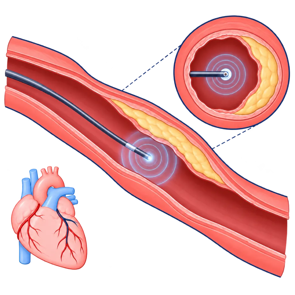
Optical coherence tomography (OCT) is a test performed from inside the coronary arteries using a thin catheter that emits infrared light. The images are very detailed, so your doctor can see the type of plaque, measure the blood vessel, choose the stent size, and make sure the stent is properly placed. OCT is performed together with cardiac catheterization.
Sources01 / Understanding the procedure
About Optical Coherence Tomography
What Is Optical Coherence Tomography
Coronary angiography shows the silhouette of the inner channel of the blood vessels from the outside, but it does not show the vessel wall itself. Optical coherence tomography (OCT) complements it by looking directly from inside the coronary artery. A very thin catheter with an infrared light source at its tip is pulled back slowly inside the artery, producing cross-sectional images of the vessel wall in very fine detail.
What It Can Show
- Type of plaque: fatty plaque, calcified plaque, or blood clots, including the thickness of the cap covering the plaque.
- Vessel size and length of the narrowing: to choose the right stent diameter and length.
- Stent placement results: whether the stent has fully expanded, sits tightly against the wall, and has not caused a tear at its edges.
- Causes of problems with an older stent: for example renarrowing or a clot inside the stent.
- Causes of certain heart attacks: for example a spontaneous tear in the coronary artery wall or plaque erosion.
How It Works in Brief
During the few seconds of image capture, blood in the artery is temporarily cleared with a flush of contrast agent, because light cannot pass through blood.
OCT and IVUS
OCT and intravascular ultrasound (IVUS) both look from inside the blood vessel. OCT gives much more detailed images, while IVUS uses sound waves that penetrate deeper and does not require a contrast flush. Your doctor chooses one of them based on the shape of the narrowing, the size of the vessel, and your kidney function. Large studies show that stent placement guided by imaging from inside the blood vessel reduces heart attacks and repeat procedures compared with angiography alone, especially in complex narrowings.
02 / Understanding the procedure
When Is This Procedure Considered?
A doctor may consider this procedure in the situations below. The decision is always made after a thorough assessment.
- Guiding Stent Placement in Complex NarrowingsFor example long narrowings, branch points, the left main artery, or heavily calcified plaque.
- Confirming Stent Placement ResultsAssessing whether the stent has fully expanded and sits tightly against the wall, then correcting it during the same procedure if needed.
- Assessing an Uncertain NarrowingWhen angiography does not show clearly enough how severe the narrowing is or what type of plaque it contains.
- Finding the Cause of Stent ProblemsIn renarrowing or a clot inside the stent, to determine the right treatment.
- Heart Attack with an Unclear CauseFor example a suspected spontaneous tear in the coronary artery wall or a heart attack without severe narrowing.
Situations to Consider First
- Severe Kidney ImpairmentOCT requires additional contrast agent. IVUS may be a more suitable option.
- Narrowing at the Artery Opening Very Close to the AortaBlood is hard to clear completely at this location, so IVUS is often preferred.
- Unstable ConditionWith very low blood pressure or a severe emergency, the main treatment takes priority.
- Contrast Agent AllergyAnti-allergy medicine can be given beforehand. Tell the team early on.
- Very Severe or Tortuous NarrowingThe OCT catheter may not be able to pass yet, so the narrowing is widened beforehand with a small balloon.
03 / Before the procedure
How Should You Prepare?
OCT is performed as part of cardiac catheterization or stent placement, so the preparation follows that procedure. The team will give you written instructions.
Tests before the procedure. Blood tests, including kidney function and blood clotting, and an ECG.
Medicines you are taking. Bring a list of all your medicines. Antiplatelet medicines are usually continued, while other blood thinners and some diabetes medicines such as metformin may need to be adjusted. Do not change your medicines without your doctor's guidance.
Fasting. Follow the fasting instructions from the team, usually for a few hours before the procedure.
Allergies. Tell the team if you have ever had an allergy to contrast agent, medicines, adhesive tape, or latex.
Drink enough. If you have no fluid restrictions, drinking enough plain water the day before helps protect your kidneys from the contrast agent.
Companion. Arrange for someone to take you home, because you are advised not to drive after the procedure.
Unsure about the preparation instructions? Ask our team before the procedure.
04 / During the procedure
How Is the Procedure Done?
The whole catheterization procedure with OCT usually takes about one hour, longer if a stent is also placed. OCT imaging itself only adds about 10 to 15 minutes.
Blood Vessel Access
After local anesthesia, your doctor inserts a small tube through an artery in your wrist or groin, then guides it to the opening of the coronary arteries. A blood thinner is given to prevent clots.
Coronary Angiography
Contrast agent is injected to show the coronary arteries and identify the section to be examined with OCT.
Inserting the OCT Catheter
A very thin guidewire is passed across the narrowing, then the OCT catheter is advanced along the wire until it is past the section to be examined.
Capturing the Images
Contrast agent is flushed for a few seconds to clear the blood, while the catheter is pulled back automatically and records images. You may feel warmth or brief discomfort in your chest.
Reading the Images and Next Steps
Your doctor assesses the images on the screen right away to determine the stent size or check the placement result. If needed, the stent is expanded again, and OCT can be repeated to confirm the result.
05 / Benefits and risks
Benefits and Risks Should Be Discussed Together.
Every medical procedure has benefits and risks. Your doctor will explain both for your situation before the procedure.
Benefits You Can Expect
- Very Detailed Images of the Vessel WallShows the type of plaque and the layers of the vessel wall that are not visible on angiography.
- More Accurate Stent SizingThe stent diameter and length are chosen based on the vessel size measured directly.
- Better Stent Placement ResultsProblems such as a stent that is not fully expanded or not fully against the wall can be corrected during the same procedure.
- Fewer Repeat EventsLarge studies show that stent placement guided by intravascular imaging reduces heart attacks and repeat procedures compared with angiography alone.
- Finding Hidden CausesHelps identify causes of a heart attack or stent problems that cannot be seen on angiography.
The benefits of a procedure are not a promise of a cure. Ask how the results will affect your care plan.
Risks You Should Know
- General Catheterization RisksBleeding or bruising at the puncture site, which is usually mild and treatable.
- Additional Contrast AgentCan affect kidney function, especially if kidney function was already reduced beforehand.
- Brief Chest DiscomfortWhile blood is cleared with contrast, blood flow to the heart muscle drops briefly. This usually goes away within seconds.
- Vessel Spasm or Rhythm DisturbancesRare and usually quickly resolved with medicines.
- Injury to the Coronary Artery WallVery rare, for example a tear or clot that needs immediate treatment, including stent placement.
Risks can differ from person to person. A full explanation and consent are given after an individual assessment.
06 / After the procedure
What Is Recovery Like?
Recovery follows the catheterization or stent placement performed at the same time.
After the procedure. If access was through the wrist, a compression band is applied for a few hours. If it was through the groin, you lie down for a few hours with your leg straight.
Hospital stay. After stent placement, patients usually stay one night. If it is only an examination without a stent, some patients can go home the same day.
Drink water. Drink enough plain water to help your kidneys clear the contrast agent, unless your doctor limits your fluids.
Medicines. If a stent was placed, take your antiplatelet medicine every day as prescribed and do not stop it without guidance from your cardiologist.
Activity. Avoid lifting heavy objects with the arm or leg used for the puncture for a few days.
Warning signs. Go to the emergency room right away or contact the medical team if you have:
- Chest pain, shortness of breath, or a cold sweat
- Growing swelling or bleeding at the puncture site
- A hand or leg that feels cold, pale, numb, or painful
- Rash, itching, or shortness of breath after receiving contrast agent
Know the Signs That Need Prompt Care.
Follow the team's guidance on warning signs. If severe or sudden symptoms appear after the procedure, go to the nearest emergency department right away or call Heart Attack Center 1500 258.
07 / Team and support
Doctors and Procedure Support
The doctors below are listed for this procedure. Listed by name, not by ranking.

dr. Adrianus Kosasih, Sp.JP(K)
Interventional Cardiology
Coronary Intervention Procedures
Clinical focusPrimary Coronary Intervention · PCI for Acute Coronary Syndrome
View Profile
Dr. dr. Dafsah A. Juzar, Sp.JP(K)
Interventional Cardiology
Coronary Intervention Procedures +1 center
Clinical focusComplex Coronary Intervention · Cardiac Catheterization
View Profile
dr. Puti S. Saus, Sp.JP(K)
Interventional Cardiology
Coronary Intervention Procedures
Clinical focusAcute Coronary Syndrome · Primary Coronary Intervention
View Profile08 / Common questions
Before You Plan the Procedure.
Frequently asked questions about optical coherence tomography. The decision to use OCT is made by your doctor when planning catheterization or stent placement.
What is the difference between OCT and coronary angiography?
Coronary angiography shows the inner channel of the blood vessels from the outside, like a silhouette. OCT looks directly from inside the blood vessel and shows its wall and plaque in great detail. For this reason, OCT is used to complement angiography, especially when planning stent placement and confirming the result.
What is the difference between OCT and IVUS?
Both look from inside the blood vessel. OCT uses infrared light and gives much more detailed images, but it requires a flush of contrast agent. IVUS uses sound waves, penetrates deeper, and does not require additional contrast. Your doctor chooses based on the location of the narrowing, the size of the vessel, and your kidney function.
Does OCT hurt?
You cannot feel the catheter moving inside the blood vessel. When contrast agent is flushed for a few seconds to capture the images, some patients feel warmth or brief discomfort in the chest. This feeling usually goes away within seconds. Tell the team if the discomfort does not go away quickly.
Does every stent placement require OCT?
Not always. Imaging from inside the blood vessel is mainly recommended for complex narrowings, for example long narrowings, branch points, the left main artery, or heavily calcified plaque. For simple narrowings, your doctor may decide that angiography is enough. Ask your doctor whether OCT or IVUS is planned for you.
How much extra time does OCT add?
OCT image capture itself takes only a few seconds for each catheter pullback, and all the steps usually add about 10 to 15 minutes to the catheterization. If OCT is repeated after the stent is placed, the time increases slightly. The length of the whole procedure is mainly determined by how complex the narrowing is.
Does OCT increase the risk to the kidneys?
OCT requires additional contrast agent to clear the blood while images are captured. With normal kidney function, this extra amount is usually safe. If kidney function is already reduced, your doctor limits the amount of contrast, gives intravenous fluids, or chooses IVUS, which does not require additional contrast. Kidney function can be rechecked after the procedure.
Sources and Medical Review
Translated from the Indonesian edition medically reviewed by dr. Puti S. Saus, Sp.JP(K).
Sources as cited in the Indonesian edition.
- Vrints C, Andreotti F, Koskinas KC, et al. 2024 ESC Guidelines for the management of chronic coronary syndromes. Eur Heart J. 2024;45(36):3415-3537.
- Ali ZA, Landmesser U, Maehara A, et al. Optical Coherence Tomography-Guided versus Angiography-Guided PCI. N Engl J Med. 2023;389(16):1466-1476.
- Holm NR, Andreasen LN, Neghabat O, et al. OCT or Angiography Guidance for PCI in Complex Bifurcation Lesions. N Engl J Med. 2023;389(16):1477-1487.
- Stone GW, Christiansen EH, Ali ZA, et al. Intravascular imaging-guided coronary drug-eluting stent implantation: an updated network meta-analysis. Lancet. 2024;403(10429):824-837.