
Quick answer
What Is Pulmonary Angiography?
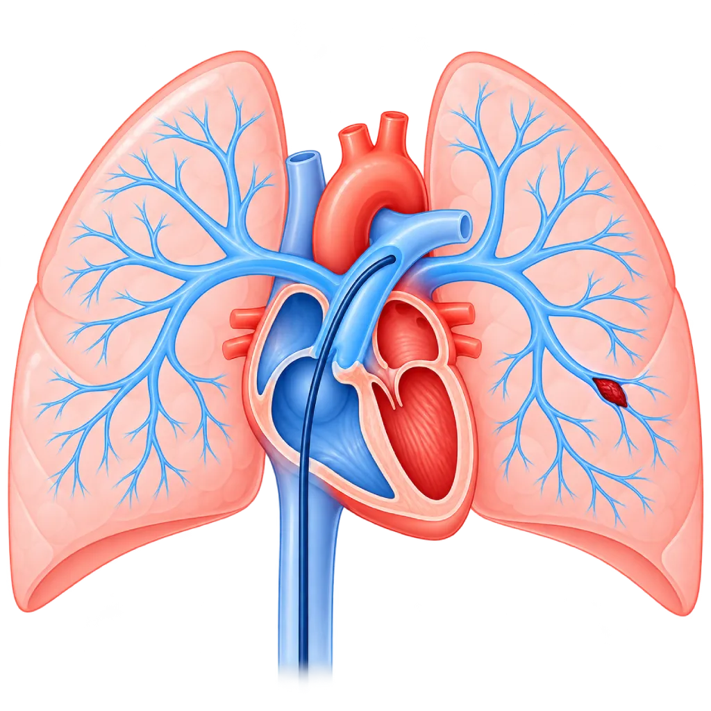
Pulmonary angiography is an examination of the pulmonary arteries in which a thin catheter is passed from a vein in the groin or neck to the right heart, then contrast dye is injected while X-ray images are recorded. It is used when CT is not enough, especially to assess long-standing blockages in pulmonary hypertension and plan its treatment.
Sources01 / Understanding the procedure
About Pulmonary Angiography
What Is Pulmonary Angiography
Pulmonary angiography is an examination to view the pulmonary arteries, the vessels that carry blood from the right side of the heart to the lungs. A thin catheter is inserted through a vein, then contrast dye is injected directly into the pulmonary artery so that its branches show up clearly on X-ray images. This examination is often done together with right heart catheterization to measure the pressures in the heart and lungs.
How It Works in Brief
The catheter enters through a vein in the groin or neck and is gently advanced through the right atrium and right ventricle into the pulmonary artery. When the contrast dye is injected, the X-ray machine records the blood flow from several angles. Parts of the artery that are blocked, narrowed, or not receiving blood appear as gaps or cutoffs in the images.
Current Uses
For suspected acute pulmonary embolism, the initial test is usually CT pulmonary angiography, which does not require a catheter. Catheter-based pulmonary angiography is now used in more specific situations:
- Chronic thromboembolic pulmonary hypertension (CTEPH): mapping long-standing blockages to decide whether pulmonary endarterectomy surgery, balloon pulmonary angioplasty, or medicines are more suitable.
- Severe pulmonary embolism: in certain situations, a catheter is used to break up or suction out the clot when clot-dissolving medicines cannot be given.
- Pulmonary artery narrowing: for example in congenital abnormalities or inflammation of the blood vessels, including before a widening procedure.
- Pulmonary arteriovenous malformation: an abnormal connection between a pulmonary artery and a pulmonary vein, which can be closed directly through the catheter.
- Unclear results from other tests: when CT or a ventilation-perfusion scan has not given an answer.
Pulmonary Angiography and the Heart
Blockages in the pulmonary arteries make the right side of the heart work harder. If this continues for a long time, the right ventricle can enlarge and weaken. That is why pulmonary angiography is often done together with pressure measurements in the right heart, so the doctor gets a picture of the lung vessels as well as the load placed on the heart.
02 / Understanding the procedure
When Is This Procedure Considered?
A doctor may consider this procedure in the situations below. The decision is always made after a thorough assessment.
- Suspected Chronic Thromboembolic Pulmonary HypertensionMapping long-standing blockages and deciding on treatment options.
- Severe Pulmonary Embolism Requiring Catheter TreatmentWhen clot-dissolving medicines cannot be given or have not worked.
- Pulmonary Artery NarrowingIn congenital abnormalities or inflammation of the blood vessels, including before a widening procedure.
- Pulmonary Arteriovenous MalformationTo confirm it and, if needed, close it directly through the catheter.
- Unclear CT or Lung Scan ResultsProvides a more detailed picture of the lung vessels.
Situations to Consider First
- Severe Allergy to Contrast DyeAllergy-preventing medicines or a different test are needed.
- Severely Reduced Kidney FunctionContrast dye can worsen kidney problems, so special preparation is needed.
- Left Bundle Branch BlockA catheter in the right ventricle can trigger a conduction disturbance, so the doctor prepares a temporary pacemaker.
- Severe Right Heart FailureThe risk is higher; the procedure is done with great care at an experienced center.
- PregnancyThe examination uses X-rays, so its benefits and risks are carefully weighed.
- Clotting Disorders or Active InfectionThe procedure is postponed until these conditions are under control, when possible.
03 / Before the procedure
How Should You Prepare?
The team will explain the preparation based on your condition.
Fasting. You are usually asked not to eat for a few hours before the procedure. Follow the fasting time given by the staff.
Blood tests. Kidney function, blood clotting, and a complete blood count are checked before the procedure.
Blood thinners. The doctor will tell you whether this medicine needs to be stopped or adjusted. Do not stop it on your own.
Other medicines. Bring a list of your medicines. If you take metformin and your kidney function is reduced, the doctor may ask you to pause it temporarily.
Important information. Tell the staff if you have ever had an allergy to contrast dye, are pregnant, or have kidney problems.
Companion. Bring a family member to accompany you and help you get home.
Unsure about the preparation instructions? Ask our team before the procedure.
04 / During the procedure
How Is the Procedure Done?
The procedure is done in the catheterization lab and usually takes about one hour. You stay awake and may be given a mild sedative.
Preparation and Local Anesthesia
The skin in the groin or neck is cleaned, then numbed with a local anesthetic so you feel only pressure, not pain.
Catheter to the Pulmonary Artery
A thin catheter is inserted into a vein and gently advanced under X-ray guidance through the right heart into the pulmonary artery. The pressures in the right heart and pulmonary artery are usually measured at this stage.
Contrast Dye and Imaging
Contrast dye is injected while the X-ray machine records from several angles. You may feel a brief warm sensation or an urge to cough, and you will be asked to hold your breath for a moment.
Finishing and Closing
The catheter is removed, then the puncture site is pressed for a few minutes and covered with a bandage.
05 / Benefits and risks
Benefits and Risks Should Be Discussed Together.
Every medical procedure has benefits and risks. Your doctor will explain both for your situation before the procedure.
Benefits You Can Expect
- A Detailed Picture of the Lung VesselsEven small branches of the pulmonary arteries can be assessed.
- Guiding CTEPH Treatment ChoicesHelps choose between surgery, balloon pulmonary angioplasty, or medicines.
- Pressure Measurement at the Same TimeTogether with right heart catheterization, the load on the right heart can be assessed.
- Can Be Followed by TreatmentIn certain situations, further treatment can be done through the same catheter.
- No Large IncisionOnly a small puncture in the groin or neck is needed.
The benefits of a procedure are not a promise of a cure. Ask how the results will affect your care plan.
Risks You Should Know
- Bruising or Bleeding at the Puncture SiteUsually mild and improves within a few days.
- Brief Rhythm DisturbancesThe catheter passing through the right heart can trigger brief palpitations; these usually stop when the catheter is repositioned.
- Reaction to Contrast DyeFrom mild itching to, very rarely, a severe reaction that the team treats right away.
- Kidney ProblemsContrast dye can worsen kidney problems in patients whose kidney function is already reduced.
- X-Ray ExposureThe dose is kept as low as possible for the needs of the examination.
- Serious ComplicationsThese are rare, but the risk is higher in very severe pulmonary hypertension.
Risks can differ from person to person. A full explanation and consent are given after an individual assessment.
06 / After the procedure
What Is Recovery Like?
After the procedure, you are monitored for a few hours in the recovery room. Some patients, especially those with severe pulmonary hypertension, stay in the hospital overnight.
Lying down. You are asked to lie down for a few hours and not to bend your leg if the catheter was inserted through the groin.
Drinking. Drinking enough water helps your kidneys clear the contrast dye, unless your doctor limits your fluids.
Activity. Avoid lifting heavy objects for a few days, as directed by your doctor.
Warning signs. Go to the emergency room right away or contact the medical team if you experience:
- Bleeding, or swelling that gets bigger, at the puncture site
- Shortness of breath or chest pain
- An irregular, pounding heartbeat or fainting
- Itching, rash, or shortness of breath after the contrast dye
- A sharp drop in the amount of urine
Know the Signs That Need Prompt Care.
Follow the team's guidance on warning signs. If severe or sudden symptoms appear after the procedure, go to the nearest emergency department right away or call Heart Attack Center 1500 258.
07 / Team and support
Doctors and Procedure Support
The doctors below are listed for this procedure. Listed by name, not by ranking.

Dr. dr. Dafsah A. Juzar, Sp.JP(K)
Interventional Cardiology
Coronary Intervention Procedures +1 center
Clinical focusComplex Coronary Intervention · Cardiac Catheterization
View Profile
dr. Emanoel Oepangat, Sp.JP(K)
Interventional Cardiology
Vascular Diagnostic and Therapeutic Center +1 center
Clinical focusCoronary Intervention · Peripheral Vascular Intervention
View Profile
dr. Santi R. Dewayanti, Sp.P
Pulmonology
Clinical focusLung and Respiratory Disease · Asthma and COPD
View Profile08 / Common questions
Before You Plan the Procedure.
Frequently asked questions about pulmonary angiography. Whether you need this procedure is decided by your doctor.
What is the difference between pulmonary angiography and CT pulmonary angiography?
CT pulmonary angiography uses a CT scan and contrast dye injected through an IV line in the arm, without a catheter, and is the initial test for acute pulmonary embolism. Pulmonary angiography uses a catheter inserted into the pulmonary artery, so the images are more detailed and pressure measurements can be done at the same time. Because it is more invasive, this procedure is used in specific situations.
Is this procedure painful?
Local anesthesia makes the puncture in the groin or neck feel only like pressure. The catheter moving inside the blood vessel usually cannot be felt. When the contrast dye is injected, you may feel a brief warm sensation or an urge to cough. If you feel anxious, the doctor can give you a mild sedative.
Do I need to stay in the hospital?
Many patients can go home on the same day after being monitored for a few hours. However, patients with severe pulmonary hypertension or kidney problems, or those who undergo further treatment, often stay overnight so the condition of the heart, kidneys, and puncture site can be monitored more closely. The doctor will tell you the plan before the procedure.
What is CTEPH and why does it require pulmonary angiography?
CTEPH is pulmonary hypertension caused by old blood clots that did not dissolve and turned into scar tissue in the pulmonary arteries. This disease can be treated with surgery, balloon pulmonary angioplasty, or medicines. Pulmonary angiography maps the location of the blockages in detail so the team can choose the appropriate treatment.
What if I have had an allergy to contrast dye before?
Tell your doctor from the start. If the allergy was mild, the doctor can give allergy-preventing medicine before the procedure and monitor you more closely. If you have had a severe reaction, the doctor will reconsider whether the procedure is needed or choose a different test. The team in the catheterization lab is also ready to handle allergic reactions.
When can I return to my activities?
The majority of patients can return to light activities the next day. Avoid lifting heavy objects and strenuous exercise for a few days so the puncture site heals well. The exact timing depends on the condition of your heart and lungs and on any further treatment performed, so follow your doctor's directions.
Sources and Medical Review
Translated from the Indonesian edition medically reviewed by dr. Santi R. Dewayanti, Sp.P.
Sources as cited in the Indonesian edition.
- Humbert M, Kovacs G, Hoeper MM, et al. 2022 ESC/ERS Guidelines for the diagnosis and treatment of pulmonary hypertension. Eur Heart J. 2022;43(38):3618-3731.
- Delcroix M, Torbicki A, Gopalan D, et al. ERS statement on chronic thromboembolic pulmonary hypertension. Eur Respir J. 2021;57(6):2002828.
- Konstantinides SV, Meyer G, Becattini C, et al. 2019 ESC Guidelines for the diagnosis and management of acute pulmonary embolism developed in collaboration with the European Respiratory Society (ERS). Eur Heart J. 2020;41(4):543-603.
- Hofmann LV, Lee DS, Gupta A, et al. Safety and hemodynamic effects of pulmonary angiography in patients with pulmonary hypertension: 10-year single-center experience. AJR Am J Roentgenol. 2004;183(3):779-785.
- Davenport MS, Perazella MA, Yee J, et al. Use of intravenous iodinated contrast media in patients with kidney disease: consensus statements from the American College of Radiology and the National Kidney Foundation. Radiology. 2020;294(3):660-668.