
Quick answer
What Is Aortic Recoarctation Repair?
Aortic recoarctation repair is a procedure to reopen an aorta that has narrowed again at the site of a previously repaired coarctation. In teenagers and adults, the repair is usually done through a catheter with a balloon and stent, without opening the chest. If the anatomy does not allow this, your doctor may recommend repeat surgery.
Sources01 / Understanding the procedure
About Aortic Recoarctation Repair
What Is Aortic Recoarctation
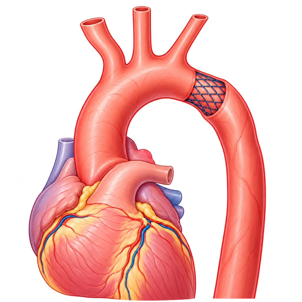
Coarctation of the aorta is a narrowing of the aorta, the main blood vessel that carries blood from the heart to the rest of the body. This narrowing is usually located just past the aortic arch, near the origin of the blood vessel to the left arm. After it has been repaired through surgery or a catheter, some patients develop narrowing again at the same site. This condition is called recoarctation.
Why the Aorta Can Narrow Again
Recoarctation is more common when the initial repair was done in infancy. The tissue around the repair site can form scar tissue or fail to grow as the aorta grows. Remnant tissue from a fetal blood vessel (the ductus arteriosus) near the narrowing can also shrink. For this reason, people with coarctation need lifelong follow-up visits even when they feel well.
Signs to Watch For
Recoarctation often causes no symptoms and is only found during a follow-up visit. Signs that may appear include high blood pressure in the arms, lower blood pressure in the legs, weak pulses in the feet, headaches, and legs that tire easily or cramp during activity. Your doctor confirms it with echocardiography, CT angiography, or MRI of the aorta.
Repair Options
The heart team chooses the repair method based on the shape and length of the narrowing, age, body size, and history of previous repairs.
- Balloon angioplasty: a balloon is inflated in the narrowed section through a catheter. It is more often used in children, because the aorta will still grow.
- Stent placement: a metal mesh tube is expanded with a balloon to hold the aorta open. In teenagers and adults, a stent is often the initial choice if the anatomy allows. A covered stent may be used to protect the aortic wall in certain situations.
- Repeat surgery: the narrowed section is repaired directly, for example with a patch or a synthetic vessel graft. This is chosen when the narrowing is long, the aortic arch is small, there is a widening at the old repair site, or another heart operation is needed at the same time.
Why Repair Is Needed
The narrowing makes the left side of the heart pump harder and raises blood pressure in the upper body. If left untreated, this can lead to thickening of the heart muscle, heart failure, widening or tearing of the aorta, and damage to the blood vessels of the brain and kidneys. Repair helps reduce this strain, although blood pressure monitoring is still needed for life.
02 / Understanding the procedure
When Is This Procedure Considered?
A doctor may consider this procedure in the situations below. The decision is always made after a thorough assessment.
- Renarrowing After Coarctation RepairThe aorta narrows again at the site of a previous surgical or catheter repair, usually found during a routine follow-up visit.
- Blood Pressure Difference Between Arms and LegsBlood pressure in the arms is more than 20 mmHg higher than in the legs, especially when accompanied by high blood pressure.
- High Blood Pressure That Is Hard to ControlHypertension in the arms, including blood pressure that rises excessively during exercise, can be a sign of significant narrowing.
- Excess Strain on the Left HeartThickening of the left heart muscle or reduced pumping function because the heart keeps working against the narrowing.
- Significant Narrowing on ImagingCT or MRI shows that the aorta is quite severely narrowed compared with the section of the aorta below it, especially when accompanied by high blood pressure.
Situations to Consider First
- Children Who Are Still SmallThe aorta will still grow, so a balloon alone, a stent that can be re-expanded later, or surgery is often preferred over a standard stent.
- Long Narrowing or Small Aortic ArchThis type of narrowing is often better suited to repair through surgery than through a catheter.
- Widening of the Aorta at the Old Repair SiteAn aneurysm at the repair site requires special planning, for example a covered stent or surgery.
- Contrast Dye Allergy or Kidney ProblemsThe procedure may still be possible with special preparation. Tell the team early on.
- Pregnant or Planning a PregnancyThe timing and method of the procedure need to be planned together with your cardiologist and obstetrician.
- Infection or FeverA planned procedure is usually postponed until the infection has cleared.
03 / Before the procedure
How Should You Prepare?
Preparation depends on the repair method chosen, through a catheter or surgery. The team will give you written instructions based on your plan.
Tests before the procedure. These usually include blood pressure measurement in the arms and legs, echocardiography, CT angiography or MRI of the aorta, an ECG, and blood tests for kidney function and blood clotting.
Medicines you are taking. Bring a list of all your medicines and supplements. Blood thinners and some diabetes medicines may need to be adjusted before the procedure, but do not stop any medicine without your doctor's guidance.
Fasting. Follow the fasting instructions from the anesthesia team, usually for several hours before the procedure. Instructions for children may differ and will be explained separately.
Allergies. Tell the team if you have ever had an allergy to contrast dye, medicines, adhesive tape, or latex.
For child patients. Explain to your child in simple words what will happen. Parents can stay with their child until the child is put to sleep, and the child may bring a favorite item.
Plan for a companion and rest time. Arrange for someone to take you home. After a catheter procedure, the hospital stay is usually 1 to 2 days; after surgery, it is longer.
Unsure about the preparation instructions? Ask our team before the procedure.
04 / During the procedure
How Is the Procedure Done?
In the majority of cases, recoarctation can be repaired through a catheter in the catheterization lab. Repeat surgery is done when the shape of the narrowing does not allow repair through a catheter.
Anesthesia and Blood Vessel Access
The procedure is usually done under general anesthesia. The doctor inserts a thin tube (catheter) through an artery in the groin, sometimes also through an artery in the arm, then guides it to the aorta using X-ray imaging.
Measuring and Mapping the Narrowing
Blood pressure is measured above and below the narrowing to assess its severity. Contrast dye is injected to show the length, diameter, and shape of the narrowing, including its relationship to the blood vessels that supply the arms.
Opening the Narrowing with a Balloon or Stent
A balloon is inflated in the narrowed section. In teenagers and adults, a stent is usually placed: it is expanded with a balloon and stays inside the aorta to hold it open. If needed, the doctor uses a covered stent.
Confirming the Result
Pressure is measured again and contrast dye is injected again to make sure the aorta is open and its wall is intact. The catheter is then removed, and the opening in the blood vessel is closed with pressure or a special closure device.
When Repeat Surgery Is Needed
Surgery is done under general anesthesia, usually through an incision on the left side of the chest, or in the middle of the chest if the aortic arch is repaired as well. The narrowed section is repaired by rejoining the aorta, with a patch, or with a synthetic vessel graft.
05 / Benefits and risks
Benefits and Risks Should Be Discussed Together.
Every medical procedure has benefits and risks. Your doctor will explain both for your situation before the procedure.
Benefits You Can Expect
- Restored Blood Flow to the Lower BodyOpening the narrowing makes it easier for blood to flow to the abdomen, kidneys, and legs.
- Helps Control Blood PressureBlood pressure in the arms often drops after repair, although some patients still need medicine.
- Reduces Strain on the HeartThe left side of the heart no longer pumps against resistance, which lowers the risk of muscle thickening and heart failure.
- Lowers the Risk of Long-Term ComplicationsHigh pressure that is left untreated can damage the blood vessels of the brain, the kidneys, and the aorta.
- An Option Without Opening the ChestMany cases of recoarctation can be repaired through a catheter, with a shorter recovery than surgery.
The benefits of a procedure are not a promise of a cure. Ask how the results will affect your care plan.
Risks You Should Know
- Bleeding or Bruising at the Puncture SiteThis is more common after a catheter procedure and is usually mild. Sometimes longer pressure or additional treatment is needed.
- Injury to the Aortic WallRare but serious: a tear in the aortic wall or a widening (aneurysm) at the site of the narrowing. The team keeps covered stents and further treatment ready in case this happens.
- Recurrent NarrowingThe aorta can narrow again, especially in children who are still growing, so the balloon or stent may need to be re-expanded later on.
- Stent Movement or FractureThis is rare and can usually be treated through a catheter.
- Contrast Reactions and Kidney ProblemsContrast dye can cause an allergic reaction or affect kidney function, especially if kidney function is already reduced.
- Risks If Surgery Is PerformedBleeding, infection, hoarseness from nerve injury, leakage of lymph fluid into the chest cavity, and, very rarely, reduced blood flow to the spinal cord.
Risks can differ from person to person. A full explanation and consent are given after an individual assessment.
06 / After the procedure
What Is Recovery Like?
Recovery time depends on the type of repair performed.
After a catheter procedure. You lie down for a few hours with your leg straight so the puncture site does not bleed. The hospital stay is usually 1 to 2 days. Light activity can be resumed within a few days, while strenuous exercise and lifting heavy objects are usually postponed for about one week or as advised.
After surgery. You are cared for in the intensive care unit initially, then on a regular ward, usually for about one week. Wound pain is controlled with medicine. Avoid lifting heavy objects for several weeks until the wound has healed.
Medicines. After stent placement, your doctor may prescribe antiplatelet medicine such as aspirin for several months. Blood pressure medicines may still be needed.
Blood pressure. Measure your blood pressure regularly at home and bring your records to your follow-up visits. Blood pressure can stay high even after the narrowing has been opened.
Follow-up visits and regular imaging. A CT or MRI of the aorta is usually done a few months after the procedure, then at regular intervals, to check the stent, the aortic wall, and any renarrowing. Follow-up with your cardiologist continues for life.
Warning signs. Go to the emergency room right away or contact the medical team if you have:
- Sudden, severe chest or back pain
- Swelling that keeps growing, or bleeding at the puncture site
- A leg that feels cold, pale, numb, or painful
- Fever, or a wound that is red, swollen, and oozing pus
- Shortness of breath, or a severe headache with very high blood pressure
Know the Signs That Need Prompt Care.
Follow the team's guidance on warning signs. If severe or sudden symptoms appear after the procedure, go to the nearest emergency department right away or call Heart Attack Center 1500 258.
07 / Team and support
Doctors and Procedure Support
The doctors below are listed for this procedure. Listed by name, not by ranking.

Dr. dr. Dudy A. Hanafy, Sp.BTKV, Subsp. JD(K), MARS
Adult Cardiac Surgery
Advanced Cardiovascular Surgical Care +1 center
Clinical focusMinimally Invasive Cardiac Surgery · Coronary Artery Bypass Surgery
View Profile
dr. Radityo Prakoso, Sp.JP(K)
Pediatrics & Congenital Heart Disease
Congenital and Structural Heart Center
Clinical focusPediatric Cardiology Consultation · Congenital Heart Disease Care
View Profile
dr. Salomo Purba, Sp.BTKV, Subsp.JPK(K)
Pediatric and Congenital Heart Surgery
Congenital and Structural Heart Center +2 centers
Clinical focusPediatric Heart Surgery · Congenital Heart Defect Surgery
View Profile08 / Common questions
Before You Plan the Procedure.
Questions patients and families often ask about aortic recoarctation repair. Your treatment plan is decided after an evaluation by the heart team.
What is aortic recoarctation?
Recoarctation is a renarrowing of the aorta at the site of a coarctation that was previously repaired, either through surgery or a catheter. It is more common when the initial repair was done in infancy, because the tissue around the repair site can form scar tissue or fail to grow as the aorta grows. For this reason, people with coarctation need lifelong follow-up visits.
Does recoarctation always need to be repaired?
Not always. Mild narrowing without high blood pressure and without strain on the heart may only need monitoring. Repair is usually recommended when the blood pressure difference between the arms and legs is more than 20 mmHg along with high blood pressure, or when imaging shows significant narrowing. The heart team makes the decision after reviewing your test results.
Is it better to repair it through a catheter or with surgery?
It depends on the shape of the narrowing and the patient's condition. In teenagers and adults, stent placement through a catheter is often the initial choice if the anatomy allows. Repeat surgery is considered when the narrowing is long, the aortic arch is small, there is a widening that cannot be treated with a stent, or another heart operation is needed at the same time.
Can my child have a stent?
Yes, depending on the size of your child's body and aorta. In young children, a balloon alone or surgery is often preferred because the aorta will still grow. In older children, the doctor may use a stent that can be expanded again as the child grows, so a repeat balloon procedure may be needed later on.
Will my blood pressure return to normal after the repair?
Many patients see their blood pressure go down, but some still have high blood pressure and still need medicine, especially if the narrowing has been present for a long time. For this reason, blood pressure needs to be monitored at home and during follow-up visits, sometimes with 24-hour monitoring or an exercise test, so treatment can be adjusted.
How long is the hospital stay, and when can I return to my usual activities?
After a catheter procedure, the hospital stay is usually 1 to 2 days and light activity can be resumed within a few days. Strenuous exercise and lifting heavy objects are usually postponed for about one week or as advised. After surgery, the hospital stay is longer, about one week, and full recovery takes several weeks.
Sources and Medical Review
Translated from the Indonesian edition medically reviewed by Dr. dr. Dudy A. Hanafy, Sp.BTKV, Subsp. JD(K), MARS.
Sources as cited in the Indonesian edition.
- Stout KK, Daniels CJ, Aboulhosn JA, et al. 2018 AHA/ACC Guideline for the Management of Adults With Congenital Heart Disease. Circulation. 2019;139(14):e698-e800.
- Baumgartner H, De Backer J, Babu-Narayan SV, et al. 2020 ESC Guidelines for the management of adult congenital heart disease. Eur Heart J. 2021;42(6):563-645.
- Feltes TF, Bacha E, Beekman RH, et al. Indications for Cardiac Catheterization and Intervention in Pediatric Cardiac Disease: A Scientific Statement From the American Heart Association. Circulation. 2011;123(22):2607-2652.
- Meadows J, Minahan M, McElhinney DB, et al. Intermediate Outcomes in the Prospective, Multicenter Coarctation of the Aorta Stent Trial (COAST). Circulation. 2015;131(19):1656-1664.