
Quick answer
What Is D-Dimer Test?
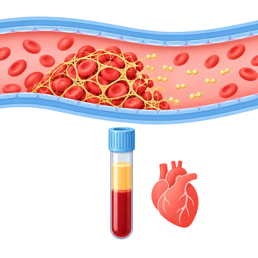
A D-dimer test measures small fragments that form when the body breaks down a blood clot. In patients with a low or moderate likelihood, a normal result helps rule out a leg vein clot and pulmonary embolism without imaging. A high result does not confirm a clot, because D-dimer also rises in many other conditions.
Sources01 / Understanding the procedure
About D-Dimer Test
What Is a D-Dimer Test
When a blood clot forms, protein strands called fibrin bind blood cells together. The body then breaks down these strands naturally, and one of the small fragments produced is called D-dimer. When a clot is forming and being broken down, the D-dimer level in the blood rises.
How It Works in Brief
Blood is drawn into a blue-capped tube containing an anticoagulant. The laboratory measures the D-dimer level in the plasma using special antibody reagents. The test is quick, so it can be used in the emergency room to help the doctor decide on the next step.
Main Use: Ruling Out a Clot
D-dimer is very sensitive but not specific for clots. Its strength therefore lies in a normal result. The doctor begins by assessing the likelihood of a clot from your symptoms and a clinical score.
- Low or moderate likelihood and normal D-dimer: deep vein thrombosis or pulmonary embolism can be ruled out without imaging.
- High D-dimer: followed by imaging, such as duplex ultrasound of the leg veins or CT pulmonary angiography.
- High likelihood: imaging is done directly, because a normal D-dimer is not enough to rule out a clot.
Result Thresholds
The commonly used threshold is 500 micrograms per liter (0.5 mg/L) in FEU units. Because D-dimer rises with age, in people over 50 many centers use an age-adjusted threshold, which is age multiplied by 10 micrograms per liter. Units and thresholds can differ between laboratories, so review your results with your doctor.
D-Dimer and the Heart
For chest pain or breathlessness that comes on suddenly, D-dimer helps the doctor assess the likelihood of pulmonary embolism. For severe chest or back pain at low risk, D-dimer is also used together with a clinical score to help rule out aortic dissection. D-dimer can rise without a clot in atrial fibrillation, heart failure, after surgery or catheterization, infection, cancer, and pregnancy, so a high result is always read together with the patient's condition.
02 / Understanding the procedure
When Is This Procedure Considered?
A doctor may consider this procedure in the situations below. The decision is always made after a thorough assessment.
- Suspected Clot in a Leg VeinSwelling, pain, and warmth in one leg.
- Suspected Pulmonary EmbolismSudden breathlessness, chest pain when breathing in, or coughing up blood.
- Sudden Severe Chest or Back PainTogether with a clinical score, helps assess pulmonary embolism or aortic dissection at low risk.
- Widespread Clotting Disorder in Severe IllnessPart of a panel to assess clotting and bleeding that occur at the same time.
- Assessing the Risk of Recurrent ClotsIn certain situations, after blood thinners have been stopped.
Situations to Consider First
- Clot Already Highly LikelyImaging is done directly, because a normal D-dimer is not enough to rule out a clot.
- PregnancyD-dimer rises naturally, so the usual threshold is often exceeded without a clot.
- Recent Surgery, Injury, or Hospital StayD-dimer is often high without a clot, so the result is less helpful.
- Infection, Cancer, or InflammationCan raise D-dimer without a clot being present.
- Currently Taking Blood ThinnersCan lower the D-dimer level, so a normal result needs to be read with caution.
- Long-Standing SymptomsIf the clot is more than one to two weeks old, the level may already have dropped.
03 / Before the procedure
How Should You Prepare?
The D-dimer test uses a blood sample and does not require fasting.
Eating and drinking. Eat and drink as usual.
Medications. Tell the staff if you are taking blood thinners, because these can affect the result. Do not stop your medication on your own.
Important information. Let the staff know if you are pregnant, have recently had surgery or an injury, have an infection, or have cancer.
Symptoms. Explain when your symptoms started, because how long they have lasted affects how the result is read.
Unsure about the preparation instructions? Ask our team before the procedure.
04 / During the procedure
How Is the Procedure Done?
The blood draw is usually completed within 10 minutes. In an emergency, this test is done quickly together with other tests in the emergency room.
Initial Assessment
The doctor asks about your symptoms and examines you, then assesses the likelihood of a clot with a clinical score before deciding whether a D-dimer test is needed.
Blood Draw
Blood is drawn from the crook of your elbow into a blue-capped tube. The tube must be filled to its mark so the ratio of blood to anticoagulant is correct.
Laboratory Testing
The plasma is separated from the blood cells, then the D-dimer level is measured and compared with the threshold used by the laboratory.
Deciding the Next Step
If the result is normal and the likelihood of a clot is low or moderate, the doctor looks for another cause. If the result is high, the doctor proceeds with imaging.
05 / Benefits and risks
Benefits and Risks Should Be Discussed Together.
Every medical procedure has benefits and risks. Your doctor will explain both for your situation before the procedure.
Benefits You Can Expect
- Fast ResultsCan be done in the emergency room to support quick decisions.
- Avoiding Unnecessary ImagingA normal result at low likelihood can spare you a CT scan with radiation and contrast.
- Helps Assess Sudden Chest PainPart of the assessment for pulmonary embolism and aortic dissection at low risk.
- Age-Adjusted ThresholdIn older people, an age-adjusted threshold reduces falsely high results.
- Just a Blood SampleNo procedure is needed other than a blood draw.
The benefits of a procedure are not a promise of a cure. Ask how the results will affect your care plan.
Risks You Should Know
- Minor Pain or BruisingAt the needle site, usually mild and quick to fade.
- High Result Without a ClotCommon in older age, pregnancy, infection, or after surgery, so further testing is needed.
- Misleading Normal ResultCan occur when taking blood thinners or when symptoms have lasted a long time.
- Infection at the Puncture SiteVery rare because the skin is cleaned and the equipment used is sterile and single-use.
Risks can differ from person to person. A full explanation and consent are given after an individual assessment.
06 / After the procedure
What Is Recovery Like?
There is no recovery period. You can return to your activities right away, unless the doctor requests further tests.
Puncture site. A small bruise is normal. If you take blood thinners, press on the puncture site a little longer.
If the result is high. The doctor may request a duplex ultrasound of the leg veins, CT pulmonary angiography, or echocardiography depending on your symptoms.
If a clot is confirmed. The doctor starts blood thinners and explains how long treatment will last.
Warning signs to watch for. Go to the emergency room right away or contact the medical team if you experience:
- Shortness of breath that comes on suddenly or gets worse
- Chest pain that worsens when you breathe in, or coughing up blood
- Swelling and pain in one leg that keeps getting worse
- Severe, tearing chest or back pain
- Fainting or a very fast heartbeat
Know the Signs That Need Prompt Care.
Follow the team's guidance on warning signs. If severe or sudden symptoms appear after the procedure, go to the nearest emergency department right away or call Heart Attack Center 1500 258.
07 / Team and support
Doctors and Procedure Support
The doctors below are listed for this procedure. Listed by name, not by ranking.

Dr. dr. Dafsah A. Juzar, Sp.JP(K)
Interventional Cardiology
Coronary Intervention Procedures +1 center
Clinical focusComplex Coronary Intervention · Cardiac Catheterization
View Profile
dr. Santi R. Dewayanti, Sp.P
Pulmonology
Clinical focusLung and Respiratory Disease · Asthma and COPD
View Profile
dr. Suko Adiarto, Sp.JP(K), Ph.D.
Interventional Cardiology
Vascular Diagnostic and Therapeutic Center +1 center
Clinical focusCoronary Intervention · Peripheral Vascular Intervention
View Profile08 / Common questions
Before You Plan the Procedure.
Frequently asked questions about the D-dimer test. Your doctor will explain what the results mean for your condition.
What does a high D-dimer mean?
A high D-dimer means clots are being formed and broken down in the body, but this is not necessarily due to a clot in a vein or the lungs. The level also rises with older age, pregnancy, infection, cancer, and after surgery. For this reason, the doctor usually follows up with imaging to confirm whether a clot is present.
Does a normal D-dimer mean I definitely have no clot?
In patients with a low or moderate likelihood of a clot, a normal D-dimer can rule out deep vein thrombosis and pulmonary embolism fairly safely. However, if the doctor judges the likelihood of a clot to be high, imaging is still done even if the D-dimer is normal. Blood thinners and long-standing symptoms can also make the result look normal.
Why is the D-dimer threshold different in older people?
D-dimer levels rise naturally with age. If the same threshold were used for all ages, many older people would appear to have high levels without a clot and would undergo unnecessary CT scans. For this reason, in people over 50, a threshold of age multiplied by 10 micrograms per liter is often used.
Can the D-dimer test be used in pregnant women?
Yes, but it is more limited. D-dimer rises naturally during pregnancy, especially toward delivery, so the result is often above the usual threshold without a clot being present. The doctor may use a special approach for pregnancy and often recommends a leg vein ultrasound directly if there are symptoms of a clot.
Can a D-dimer test detect a heart attack?
No. A heart attack is assessed with an ECG and a troponin test. D-dimer is used to help assess clots in the veins and lungs, as well as aortic dissection in low-risk patients. For sudden chest pain, doctors often run several tests at once to find the right cause.
I take blood thinners. Will this affect my result?
It can. Blood thinners help prevent new clots, so the D-dimer level may be lower than it would otherwise be. For this reason, a normal result in people taking these medications needs to be read with caution. Tell your doctor which medication you take and when you last took it, and do not stop it on your own.
Sources and Medical Review
Translated from the Indonesian edition medically reviewed by dr. Suko Adiarto, Sp.JP(K), Ph.D..
Sources as cited in the Indonesian edition.
- Konstantinides SV, Meyer G, Becattini C, et al. 2019 ESC Guidelines for the diagnosis and management of acute pulmonary embolism developed in collaboration with the European Respiratory Society (ERS). Eur Heart J. 2020;41(4):543-603.
- Lim W, Le Gal G, Bates SM, et al. American Society of Hematology 2018 guidelines for management of venous thromboembolism: diagnosis of venous thromboembolism. Blood Adv. 2018;2(22):3226-3256.
- Righini M, Van Es J, Den Exter PL, et al. Age-adjusted D-dimer cutoff levels to rule out pulmonary embolism: the ADJUST-PE study. JAMA. 2014;311(11):1117-1124.
- van der Hulle T, Cheung WY, Kooij S, et al. Simplified diagnostic management of suspected pulmonary embolism (the YEARS study): a prospective, multicentre, cohort study. Lancet. 2017;390(10091):289-297.
- Nazerian P, Mueller C, Soeiro AM, et al. Diagnostic accuracy of the aortic dissection detection risk score plus D-dimer for acute aortic syndromes: the ADvISED prospective multicenter study. Circulation. 2018;137(3):250-258.
- Weitz JI, Fredenburgh JC, Eikelboom JW. A test in context: D-dimer. J Am Coll Cardiol. 2017;70(19):2411-2420.