
Quick answer
What Is Ventilation-Perfusion Scan?
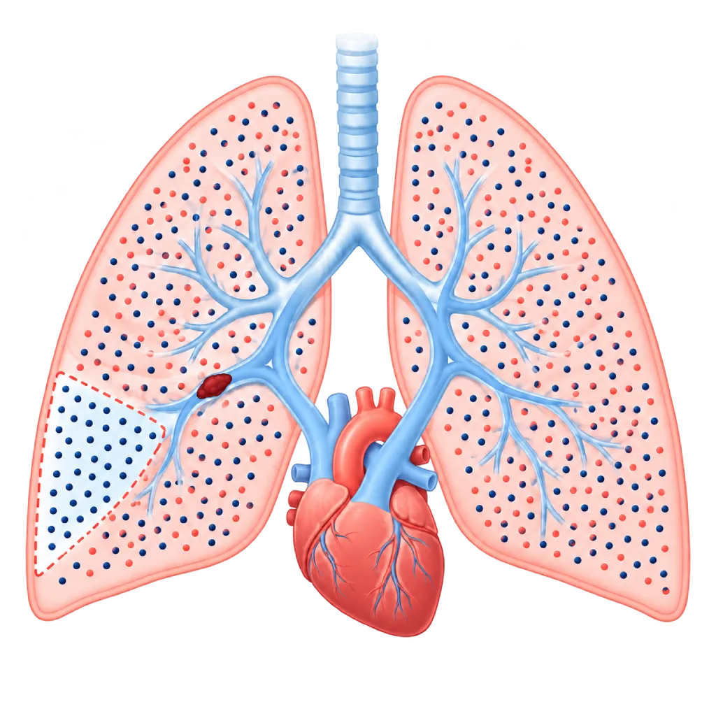
A ventilation-perfusion scan, or V/Q scan, is a nuclear medicine test that compares airflow and blood flow in the lungs. You breathe in a tracer, then receive an injection of another tracer, and a special camera takes pictures of your lungs. Areas that receive air but no blood may indicate a blockage, such as a pulmonary embolism.
Sources01 / Understanding the procedure
About Ventilation-Perfusion Scan
What Is a Ventilation-Perfusion Scan
A ventilation-perfusion scan (V/Q scan) is a nuclear medicine test for the lungs. Ventilation means airflow to the air sacs of the lungs, while perfusion means blood flow in the small blood vessels of the lungs. In healthy lungs, both are evenly distributed and match each other.
How It Works in Brief
For the ventilation images, you breathe in a mist or gas containing a low-dose radioactive tracer. For the perfusion images, very small albumin particles carrying a tracer are injected into a vein in your arm, then temporarily lodge in a small portion of the lung capillaries. A gamma camera captures the signals from both. If part of the lung receives air but no blood, this is called a mismatch and points to a blockage in a blood vessel.
When This Test Is Chosen
- Suspected pulmonary embolism: especially when CT pulmonary angiography is less suitable, for example because of an allergy to iodine contrast, severe kidney impairment, pregnancy, or young age.
- Pulmonary hypertension: to look for long-standing blood clot blockages (CTEPH), because this test is more sensitive than CT in finding them.
- Persistent shortness of breath after a pulmonary embolism: to see whether any blockage remains after several months of treatment.
Reading the Results
A normal perfusion result almost rules out a pulmonary embolism, while a widespread mismatch pattern supports a diagnosis of embolism. In some patients, especially those with other lung diseases such as COPD or pneumonia, the results may be inconclusive, so the doctor supplements them with CT pulmonary angiography. If the results point to CTEPH, further tests such as pulmonary angiography and right heart catheterization may be needed.
02 / Understanding the procedure
When Is This Procedure Considered?
A doctor may consider this procedure in the situations below. The decision is always made after a thorough assessment.
- Suspected Pulmonary EmbolismEspecially if CT with iodine contrast is less suitable for you.
- Contrast Allergy or Kidney ProblemsThis test does not use iodine contrast.
- Suspected Pulmonary Embolism During PregnancyCan be done with a reduced dose of tracer.
- Pulmonary HypertensionTo look for long-standing blockages (CTEPH) that can be treated.
- Persistent Shortness of Breath After a Pulmonary EmbolismAssesses remaining blockages after several months of treatment.
- Monitoring Treatment ResultsCompares lung blood flow before and after therapy.
Situations to Consider First
- PregnancyCan still be done if needed. Tell the staff so the dose can be adjusted.
- BreastfeedingBreastfeeding may need to be paused for a few hours according to the staff's instructions.
- Severe Pulmonary Hypertension or a Heart ShuntThe number of particles injected is reduced for safety.
- Difficulty Lying StillImages may be blurred and need to be repeated.
03 / Before the procedure
How Should You Prepare?
This test generally does not require fasting.
Chest X-ray. Bring a recent chest X-ray if you have one, because the doctor usually needs it to read the results.
Pregnancy and breastfeeding. Tell the staff if you are pregnant, might be pregnant, or are breastfeeding.
Medicines and history. Tell the staff about the medicines you take, any allergies, and any lung or heart conditions you have.
Clothing. Remove jewelry and metal objects from your chest area before the images are taken.
Unsure about the preparation instructions? Ask our team before the procedure.
04 / During the procedure
How Is the Procedure Done?
The whole test usually takes about 1 hour. The order of the ventilation and perfusion parts may differ between hospitals.
Breathing in the Tracer
You breathe normally through a mouthpiece with a nose clip for a few minutes to inhale a mist or gas containing the tracer.
Ventilation Images
You lie still while a gamma camera around your chest takes pictures of how air is distributed in your lungs.
Injection in the Arm
The second tracer is injected into a vein in your arm while you lie down. It feels like a regular injection.
Perfusion Images and Reading
The camera again takes pictures of your lungs from several angles. The doctor compares the two sets of images, then matches them with your chest X-ray and symptoms.
05 / Benefits and risks
Benefits and Risks Should Be Discussed Together.
Every medical procedure has benefits and risks. Your doctor will explain both for your situation before the procedure.
Benefits You Can Expect
- No Iodine ContrastAn option if you are allergic to contrast or have impaired kidney function.
- A Normal Result Almost Rules Out Pulmonary EmbolismNormal lung perfusion greatly helps the doctor make decisions.
- Sensitive for Detecting CTEPHMore sensitive than CT for finding long-standing blockages.
- Low Radiation Dose to the BreastsMuch lower than CT pulmonary angiography.
- Assessing Lung FunctionShows how air and blood are distributed throughout the lungs.
The benefits of a procedure are not a promise of a cure. Ask how the results will affect your care plan.
Risks You Should Know
- Low Radiation ExposureThe dose is about 2 mSv, and the tracer decays quickly.
- Inconclusive ResultsEspecially if you have another lung disease, so a CT may be needed.
- Allergic ReactionVery rare.
- Bruising at the Injection SiteMild and goes away on its own.
Risks can differ from person to person. A full explanation and consent are given after an individual assessment.
06 / After the procedure
What Is Recovery Like?
You can go home right away and resume your usual activities. The tracer decays quickly, and the majority of it leaves the body through the urine within a day.
After the test. Drink plenty of fluids and urinate regularly. If you are breastfeeding or caring for a baby, follow the staff's instructions.
Warning signs to watch for. Go to the emergency room right away or contact the medical team if you experience:
- Shortness of breath that gets worse or comes on suddenly
- Chest pain, especially when breathing in
- Coughing up blood
- Fainting, palpitations, or a swollen and painful leg
Know the Signs That Need Prompt Care.
Follow the team's guidance on warning signs. If severe or sudden symptoms appear after the procedure, go to the nearest emergency department right away or call Heart Attack Center 1500 258.
07 / Team and support
Doctors and Procedure Support
The doctors below are listed for this procedure. Listed by name, not by ranking.

Dr. med. dr. Denio A. Ridjab, Sp.JP(K)
Interventional Cardiology
Coronary Intervention Procedures +2 centers
Clinical focusInterventional Cardiology · Coronary Artery Disease
View Profile
dr. Emanoel Oepangat, Sp.JP(K)
Interventional Cardiology
Vascular Diagnostic and Therapeutic Center +1 center
Clinical focusCoronary Intervention · Peripheral Vascular Intervention
View Profile
dr. Santi R. Dewayanti, Sp.P
Pulmonology
Clinical focusLung and Respiratory Disease · Asthma and COPD
View Profile08 / Common questions
Before You Plan the Procedure.
Frequently asked questions about the ventilation-perfusion scan. Your doctor will explain what the results mean for your condition.
Is the radioactive substance in this test dangerous?
The radiation dose is low, about 2 mSv, roughly equal to natural background radiation over a few months. The tracer used decays within hours, and the majority of it leaves the body through the urine within a day. The injected particles also break down on their own. The risk is very small compared with the benefit of finding or ruling out a blockage in the lungs.
How is it different from CT pulmonary angiography?
CT pulmonary angiography uses iodine contrast and shows clots directly in the pulmonary arteries, so it is the preferred initial test for the majority of patients with suspected pulmonary embolism. A ventilation-perfusion scan assesses function, does not use contrast, and gives a lower dose to the breasts. It is also more sensitive for finding CTEPH. Your doctor chooses the test that suits your condition.
Can pregnant women have this test?
Yes, if it is truly needed to look for a pulmonary embolism, which is dangerous if left untreated. The tracer dose is usually reduced, and in some patients only the perfusion part is done if the chest X-ray is normal. Radiation exposure to the unborn baby is very low. Tell the staff early on if you are pregnant or might be pregnant.
Will the injected particles block my lungs?
Not in any meaningful way. The albumin particles only lodge temporarily in a very small portion of the lung capillaries, then break down and are cleared by the body within a few hours. You will not feel short of breath because of them. In patients with severe pulmonary hypertension or certain heart abnormalities, the number of particles is reduced to keep the test safe.
Why have I been asked to have this test when I have already had a CT?
In pulmonary hypertension, your doctor needs to confirm whether the cause is long-standing blood clot blockages (CTEPH), because this condition can be treated. To look for it, a ventilation-perfusion scan is more sensitive than CT. This test is also used when CT results are unclear. The two tests complement each other rather than replace each other.
What happens if the results show a blockage?
Your doctor will explain the results together with your symptoms and other tests. A new pulmonary embolism is usually treated with blood-thinning medicines (anticoagulants). If the results point to CTEPH, your doctor may request further tests such as pulmonary angiography and right heart catheterization to decide on treatment options, for example surgery, a balloon procedure, or medicines.
Sources and Medical Review
Translated from the Indonesian edition medically reviewed by dr. Santi R. Dewayanti, Sp.P.
Sources as cited in the Indonesian edition.
- Konstantinides SV, Meyer G, Becattini C, et al. 2019 ESC Guidelines for the diagnosis and management of acute pulmonary embolism developed in collaboration with the European Respiratory Society (ERS). Eur Heart J. 2020;41(4):543-603.
- Humbert M, Kovacs G, Hoeper MM, et al. 2022 ESC/ERS Guidelines for the diagnosis and treatment of pulmonary hypertension. Eur Heart J. 2022;43(38):3618-3731.
- Bajc M, Schümichen C, Grüning T, et al. EANM guideline for ventilation/perfusion single-photon emission computed tomography (SPECT) for diagnosis of pulmonary embolism and beyond. Eur J Nucl Med Mol Imaging. 2019;46(12):2429-2451.
- Tunariu N, Gibbs SJR, Win Z, et al. Ventilation-perfusion scintigraphy is more sensitive than multidetector CTPA in detecting chronic thromboembolic pulmonary disease as a treatable cause of pulmonary hypertension. J Nucl Med. 2007;48(5):680-684.
- Mettler FA Jr, Huda W, Yoshizumi TT, Mahesh M. Effective doses in radiology and diagnostic nuclear medicine: a catalog. Radiology. 2008;248(1):254-263.