
Quick answer
What Is Aortic Angiography?
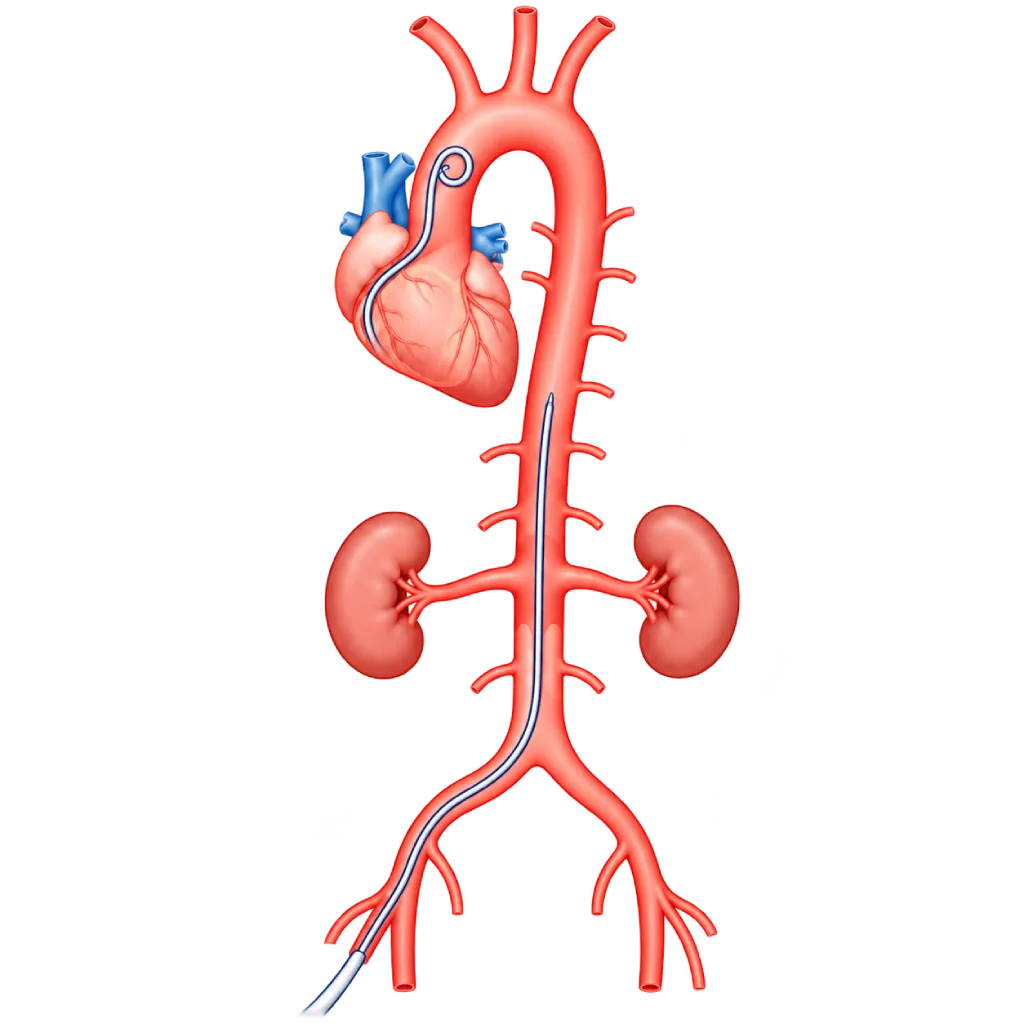
Aortic angiography is an X-ray examination of the aorta after contrast dye is injected directly into it through a catheter from a blood vessel in the groin or arm. It is used when CT or MRI is not clear enough, when pressure inside the aorta needs to be measured, or as part of a procedure on the aorta.
Sources01 / Understanding the procedure
About Aortic Angiography
What Is Aortic Angiography
Aortic angiography, also called aortography, is an examination to view the aorta and its branches with X-rays. Contrast dye is injected directly into the aorta through a catheter, so the shape of the inside of the aorta shows clearly on the screen. During the examination, the doctor can also measure blood pressure directly inside the aorta.
What Can Be Assessed
- Narrowing: of the aorta or its branches, for example coarctation of the aorta or narrowing of the kidney arteries.
- Widening (aneurysm): including its position relative to important branches such as the kidney arteries and the blood vessels to the arms.
- Tears in the wall (dissection): as well as leaks around a stent graft after it has been placed.
- Aortic valve leakage: by injecting contrast at the root of the aorta during cardiac catheterization.
- Pressure inside the aorta: including the pressure difference above and below a narrowing.
Aortic Angiography, CT Angiography, and MRI
CT angiography and MRI can show the aorta without a catheter, so they are usually the initial tests. Catheter-based aortic angiography is chosen when the results of those tests are not clear enough, when pressure inside the aorta needs to be measured directly, or when the examination is done together with a procedure, for example placing a stent graft or a coarctation stent. The doctor will explain why this examination has been chosen for you.
02 / Understanding the procedure
When Is This Procedure Considered?
A doctor may consider this procedure in the situations below. The decision is always made after a thorough assessment.
- CT or MRI Results Not Clear EnoughIf the images of the aorta or its branches are still uncertain, angiography provides a more detailed view.
- Need to Measure Pressure Inside the AortaFor example, to assess the severity of aortic coarctation from the pressure difference above and below the narrowing.
- Planning or Part of a ProcedureBefore or during placement of a stent graft, a coarctation stent, or other procedures on the aorta and its branches.
- Assessment After Stent Graft PlacementTo assess a leak around the stent graft (endoleak) seen on CT that may require treatment.
- Assessing Aortic Valve LeakageContrast is injected at the root of the aorta during cardiac catheterization when echocardiography or MRI results are not clear enough.
Situations to Consider First
- Reduced Kidney FunctionContrast dye can worsen kidney problems. The team may give IV fluids, limit the amount of contrast, or consider another test.
- Contrast Dye AllergyAnti-allergy medication can be given before the examination. Tell the team if you have had a reaction before.
- PregnancyX-ray examinations are usually postponed or replaced with another test. Tell the team if you are pregnant or might be pregnant.
- Blood Clotting Disorders or Taking Blood ThinnersMedication adjustments and care of the puncture site need to be planned in advance.
- Infection or FeverA planned examination is usually postponed until the infection has cleared.
03 / Before the procedure
How Should You Prepare?
The team will give you written instructions before the examination.
Blood tests and previous results. Kidney function and blood clotting are checked first. Bring the results of any previous CT, MRI, or echocardiography.
Medications you are taking. Bring a list of all your medications and supplements. Blood thinners and some diabetes medications, such as metformin, may need to be adjusted. Do not stop any medication without your doctor's direction.
Fasting. Usually for a few hours before the examination, as instructed by the team.
Allergies. Tell the team if you have ever been allergic to contrast dye, medications, adhesive tape, or latex.
Pregnancy. Tell the team if you are pregnant or might be pregnant.
Companion. Arrange for someone to take you home, because you are advised not to drive on the day of the examination.
Unsure about the preparation instructions? Ask our team before the procedure.
04 / During the procedure
How Is the Procedure Done?
The examination is done in the catheterization room. How long it takes depends on which part of the aorta is examined and whether pressure measurements are needed.
Preparation in the Procedure Room
You lie on the examination table and are connected to monitoring equipment. A mild sedative can be given if needed. The groin or wrist area is cleaned and then numbed with a local anesthetic.
Inserting the Catheter
The doctor inserts a thin tube (catheter) through an artery in the groin, sometimes through an artery in the arm, and guides it to the aorta using X-ray guidance. You will not feel the catheter moving inside the blood vessels.
Injecting the Contrast Dye
The contrast dye is injected quickly while X-ray images are recorded. You may feel warm all over your body for a few seconds and be asked to hold your breath briefly.
Measuring Pressure
If needed, blood pressure is measured at several points in the aorta, for example above and below a narrowing.
Removing the Catheter
Once the necessary images have been collected, the catheter is removed, and the puncture site is pressed or closed with a vascular closure device.
05 / Benefits and risks
Benefits and Risks Should Be Discussed Together.
Every medical procedure has benefits and risks. Your doctor will explain both for your situation before the procedure.
Benefits You Can Expect
- A Detailed View of the AortaClearly shows the inside of the aorta and its branches, including narrowing, widening, or tears.
- Direct Pressure MeasurementThe pressure difference inside the aorta can be measured, something CT or MRI cannot do.
- Helps Choose the Right TreatmentThe results help the team decide whether a stent, a stent graft, surgery, or simply monitoring is needed.
- Can Be Followed by a ProcedureIf needed and already planned with you, repair can be done in the same session.
- Usually No Overnight StayThe majority of patients can go home on the same day after monitoring.
The benefits of a procedure are not a promise of a cure. Ask how the results will affect your care plan.
Risks You Should Know
- Bruising or Bleeding at the Puncture SiteCommon and usually mild.
- Reaction to Contrast DyeRanges from itching or a rash to, very rarely, a severe allergic reaction that is treated right away by the team.
- Kidney ProblemsMore likely if kidney function was already reduced before the examination.
- Blood Vessel InjuryRare: a tear in the vessel wall, a blood clot, or dislodged plaque that can block blood flow to the legs, kidneys, or brain.
- X-Ray ExposureThe X-ray dose is kept as low as possible for the needs of the examination.
Risks can differ from person to person. A full explanation and consent are given after an individual assessment.
06 / After the procedure
What Is Recovery Like?
The majority of patients can go home on the same day after a few hours of monitoring.
After the examination. If the catheter was inserted through the groin, you lie down for a few hours with your leg straight. If it was inserted through the wrist, a compression band is placed for a few hours.
Drink water. Drink enough plain water to help your kidneys clear the contrast dye, unless your doctor limits your fluids.
Activity. Avoid lifting heavy objects and strenuous exercise for a few days, and do not drive on the day of the examination.
Results. The doctor explains the findings and the next steps, for example medication, a procedure, or regular monitoring.
Warning signs to watch for. Contact the medical team or go to the emergency room right away if you experience:
- Swelling that grows or bleeding at the puncture site
- A leg or arm that feels cold, pale, numb, or painful
- A rash, itching, or shortness of breath after the examination
- Severe chest, back, or abdominal pain
- A much smaller amount of urine than usual
Know the Signs That Need Prompt Care.
Follow the team's guidance on warning signs. If severe or sudden symptoms appear after the procedure, go to the nearest emergency department right away or call Heart Attack Center 1500 258.
07 / Team and support
Doctors and Procedure Support
The doctors below are listed for this procedure. Listed by name, not by ranking.

dr. Emanoel Oepangat, Sp.JP(K)
Interventional Cardiology
Vascular Diagnostic and Therapeutic Center +1 center
Clinical focusCoronary Intervention · Peripheral Vascular Intervention
View Profile
dr. Suko Adiarto, Sp.JP(K), Ph.D.
Interventional Cardiology
Vascular Diagnostic and Therapeutic Center +1 center
Clinical focusCoronary Intervention · Peripheral Vascular Intervention
View ProfileVascular Interventional Suite
A procedure room with X-ray imaging for diagnosing and treating narrowed, blocked, or enlarged blood vessels outside the heart…
The availability of technology and test support is confirmed with the team. Technology is chosen according to clinical need.
08 / Common questions
Before You Plan the Procedure.
Frequently asked questions before aortic angiography. Instructions specific to your examination are given by the team.
What is the difference between aortic angiography and CT angiography?
Both use contrast dye and X-rays to view the aorta. CT angiography is done from outside the body with contrast injected through an IV line in the arm, so no catheter is needed. Aortic angiography uses a catheter inserted into the aorta, so pressure inside the aorta can be measured directly and a procedure can follow if needed.
Is aortic angiography painful?
You will feel a prick when the local anesthetic is injected, then pressure when the catheter is inserted. You cannot feel the catheter moving inside the blood vessels. When the contrast dye is injected, your body feels warm for a few seconds. A mild sedative can be given if you feel anxious.
How long does the examination take, and do I need to stay overnight?
The length of the examination depends on which part of the aorta is examined and whether pressure measurements are needed. Afterward, you are monitored for a few hours. The majority of patients can go home on the same day, but the doctor may ask you to stay overnight in certain situations or if a procedure follows.
Is contrast dye harmful to the kidneys?
In the majority of people, the kidneys clear contrast dye without any problems. The risk of kidney problems is higher if kidney function is already reduced, in people with diabetes, or when the body is dehydrated. That is why kidney function is checked first, and the team may give IV fluids and limit the amount of contrast.
When will the results be known?
The images can be seen directly during the examination, so the doctor can usually explain the main findings on the same day. A complete report is prepared after all the images and pressure measurements have been analyzed, and it is discussed at your follow-up visit along with a suitable treatment plan, for example medication, a procedure, or monitoring.
When can I return to my usual activities?
Light activities are usually allowed the next day. Avoid lifting heavy objects and strenuous exercise for a few days so the puncture site can heal, and do not drive on the day of the examination. Follow the team's advice, especially if the catheter was inserted through the groin, and ask when you can return to work.
Sources and Medical Review
Translated from the Indonesian edition medically reviewed by dr. Suko Adiarto, Sp.JP(K), Ph.D..
Sources as cited in the Indonesian edition.
- Isselbacher EM, Preventza O, Hamilton Black J, et al. 2022 ACC/AHA Guideline for the Diagnosis and Management of Aortic Disease. Circulation. 2022;146(24):e334-e482.
- Mazzolai L, Teixido-Tura G, Lanzi S, et al. 2024 ESC Guidelines for the management of peripheral arterial and aortic diseases. Eur Heart J. 2024;45(36):3538-3700.
- Otto CM, Nishimura RA, Bonow RO, et al. 2020 ACC/AHA Guideline for the Management of Patients With Valvular Heart Disease. Circulation. 2021;143(5):e72-e227.
- Davenport MS, Perazella MA, Yee J, et al. Use of Intravenous Iodinated Contrast Media in Patients with Kidney Disease: Consensus Statements from the American College of Radiology and the National Kidney Foundation. Radiology. 2020;294(3):660-668.