
Quick answer
What Is Vena Cava Filter Placement?
Vena cava filter placement inserts a small metal filter into the large abdominal vein through a catheter to keep leg blood clots from blocking the lungs. It is used when blood thinners cannot be given or fail to prevent embolism. It takes about 1 hour under local anesthesia, and a temporary filter is removed once no longer needed.
Sources01 / Understanding the procedure
About Vena Cava Filter Placement
What Is a Vena Cava Filter
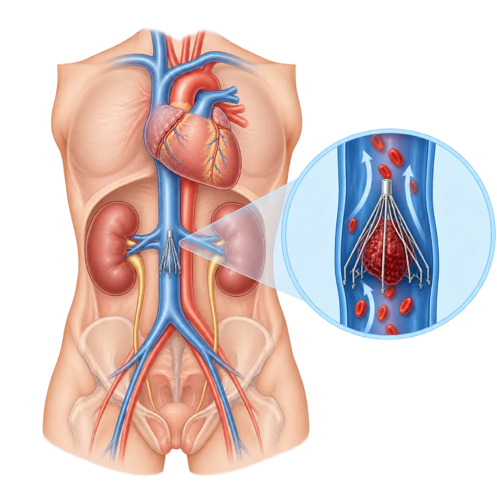
A vena cava filter is a small metal filter shaped somewhat like an umbrella with thin legs. It is placed in the inferior vena cava, the large vein in the abdomen that carries blood from the lower body to the heart, usually just below where the kidney veins join it. Its purpose is to catch blood clots that break loose from the veins of the legs or pelvis so they do not travel to the lungs and cause a pulmonary embolism.
How It Works at a Glance
Through a small puncture in a vein in the groin or neck, the doctor inserts a thin catheter using X-ray guidance. The folded filter inside the catheter is then released, so its legs open and attach to the vein wall. Blood keeps flowing through the gaps in the filter, while sufficiently large clots are trapped in it.
When a Filter Is Considered
The main treatment for deep vein thrombosis and pulmonary embolism is blood thinners, covered on the Anticoagulation Therapy and Stroke Prevention page. A filter does not treat clots and does not prevent new clots from forming. Guidelines recommend considering a filter when:
- Blood thinners cannot be given, for example because of active bleeding or a very high risk of bleeding.
- Pulmonary embolism still occurs even though blood thinners have been given at the correct dose.
Filters are not recommended for routine use in all patients, because studies have shown that adding a filter to blood thinners does not lower the risk of repeat pulmonary embolism.
Temporary Filters and Their Removal
The majority of filters used today can be removed again. Once blood thinners can be given safely, the filter should be removed, because a filter left in place for a long time is harder to remove and can increase the risk of clots in the leg veins. Removal is done in a similar way, usually through a vein in the neck.
Tests Beforehand
Clots in the leg veins are confirmed with ultrasound, covered on the Duplex Ultrasound page. Supporting blood tests for a suspected clot are covered on the D-Dimer Test page.
02 / Understanding the procedure
When Is This Procedure Considered?
A doctor may consider this procedure in the situations below. The decision is always made after a thorough assessment.
- Unable to Take Blood ThinnersIn deep vein clots or pulmonary embolism, for example because of active bleeding.
- Repeat Pulmonary Embolism Despite TreatmentBlood thinners have already been given at the correct dose.
- Blood Thinners Must Be Stopped TemporarilyFor example, before urgent major surgery soon after a new clot.
- Other Special SituationsDecided by the medical team based on each patient's risk.
Situations to Consider First
- No Usable Venous AccessIf the neck and groin veins are blocked by clots.
- Blocked or Abnormally Shaped Vena CavaThe size and shape of the vein are assessed beforehand.
- Severe Contrast AllergySpecial preparation or another method is needed.
- Uncontrolled Bloodstream InfectionPlacement is postponed when possible.
- Blood Thinners Can Be Given SafelyA filter is usually not needed.
03 / Before the procedure
How Should You Prepare?
The majority of patients who need a filter are already in the hospital, so preparation is arranged by the team on the ward.
Fasting. You usually fast for a few hours before the procedure, especially if sedation is planned. Follow the team's instructions.
Blood tests. Kidney function and blood clotting are checked because the procedure uses contrast and a puncture in a vein.
Allergy history. Tell the team if you have ever been allergic to contrast, anesthetics, or certain metals.
Medication list. Share all the medications you take, especially blood thinners and diabetes medications.
Pregnancy. Tell the team if you are or might be pregnant.
Unsure about the preparation instructions? Ask our team before the procedure.
04 / During the procedure
How Is the Procedure Done?
The procedure is done in the catheterization or angiography lab and takes about 30 minutes to 1 hour, under local anesthesia. Mild sedation can be given if needed.
Vein Access
The skin in the groin or neck is numbed, then a needle is inserted into the vein with ultrasound guidance and a small sheath is placed.
Mapping the Vena Cava
Contrast is injected to see the size of the vena cava and the position of the kidney veins, and to make sure there is no clot where the filter will be placed.
Placing the Filter
The folded filter is pushed through the catheter to just below where the kidney veins join, then released so its legs open and attach to the vein wall. Its position is checked with X-ray.
Finishing Up
The catheter is removed and the puncture site is pressed until the bleeding stops. You will be asked to lie down for a few hours.
05 / Benefits and risks
Benefits and Risks Should Be Discussed Together.
Every medical procedure has benefits and risks. Your doctor will explain both for your situation before the procedure.
Benefits You Can Expect
- Protection from Pulmonary EmbolismWhile blood thinners cannot yet be given.
- No Open SurgeryDone through a small puncture under local anesthesia.
- Can Be Removed LaterA temporary filter is removed once it is no longer needed.
- Short ProcedureUsually about 30 minutes to 1 hour.
The benefits of a procedure are not a promise of a cure. Ask how the results will affect your care plan.
Risks You Should Know
- Bruising or Bleeding at the Puncture SiteUsually mild and stops with pressure.
- Reaction to ContrastAllergy or reduced kidney function; monitored by the team.
- Filter Shifting or TiltingThis can make the filter difficult to remove.
- Filter Legs Piercing the Vein Wall or BreakingThis rarely causes symptoms, but it is more common when the filter is left in place for a long time.
- Clots in the Filter or Leg VeinsThe long-term risk of leg vein clots increases, which is why the filter is removed once it is no longer needed.
- Filter Cannot Be RemovedThis sometimes happens, especially when the filter has been in place for a long time.
Risks can differ from person to person. A full explanation and consent are given after an individual assessment.
06 / After the procedure
What Is Recovery Like?
Recovery from the procedure itself is usually quick. The length of the hospital stay depends more on the underlying condition.
Puncture site. Keep it dry and clean. A small bruise is normal.
Blood thinners. Your doctor will start or continue blood thinners as soon as it is safe, because a filter does not replace treatment of the clot.
Removal plan. If your filter can be removed, ask about the removal schedule and make sure you do not miss your follow-up visit.
Filter record. Keep a record of your filter type. Tell the staff about it before an MRI or other procedures.
Warning signs. Go to the emergency room right away if you have:
- Sudden shortness of breath or chest pain
- Sudden swelling in both legs
- Bleeding from the puncture site that does not stop with pressure
- Severe pain in the abdomen or back
Know the Signs That Need Prompt Care.
Follow the team's guidance on warning signs. If severe or sudden symptoms appear after the procedure, go to the nearest emergency department right away or call Heart Attack Center 1500 258.
07 / Team and support
Doctors and Procedure Support
The doctors below are listed for this procedure. Listed by name, not by ranking.

dr. Emanoel Oepangat, Sp.JP(K)
Interventional Cardiology
Vascular Diagnostic and Therapeutic Center +1 center
Clinical focusCoronary Intervention · Peripheral Vascular Intervention
View Profile
dr. Santi R. Dewayanti, Sp.P
Pulmonology
Clinical focusLung and Respiratory Disease · Asthma and COPD
View Profile
dr. Suko Adiarto, Sp.JP(K), Ph.D.
Interventional Cardiology
Vascular Diagnostic and Therapeutic Center +1 center
Clinical focusCoronary Intervention · Peripheral Vascular Intervention
View ProfileVascular Interventional Suite
A procedure room with X-ray imaging for diagnosing and treating narrowed, blocked, or enlarged blood vessels outside the heart…
The availability of technology and test support is confirmed with the team. Technology is chosen according to clinical need.
08 / Common questions
Before You Plan the Procedure.
Frequently asked questions about vena cava filters. The decision to place a filter is made by the medical team based on your condition.
Does a filter replace blood thinners?
No. A filter only catches large clots so they do not reach the lungs. It does not dissolve existing clots and does not prevent new clots from forming. For this reason, blood thinners remain the main treatment and will be started or continued as soon as they can be given safely, as decided by your doctor.
Does the filter have to be removed?
If your filter is a removable type and blood thinners can now be given safely, the filter should be removed. A filter left in place for a long time is harder to remove and more often causes problems, such as clots in the leg veins or filter legs piercing the vein wall. Your doctor will decide when to remove it.
How is the filter removed?
The method is similar to placement. Under local anesthesia, a catheter is inserted through a vein in the neck or groin. The tip of the filter is caught with a device like a small snare, then the filter is folded back into a sheath and pulled out. The procedure is usually short, and the majority of patients can go home the same day.
Will I be able to feel the filter inside my body?
Usually not. The filter sits inside a large vein in the abdomen and cannot be felt during daily activities. You can still walk, work, and go about your usual activities after recovering from the procedure. If you develop severe abdominal or back pain, or swelling in both legs, see a doctor right away.
Is it safe to have an MRI with a filter?
The majority of filters used today are safe for MRI under certain conditions. For this reason, keep a record of the type of filter you have and tell the staff before an MRI. The staff will check whether the scan can be done and which machine settings need to be used.
Do all patients with vein clots need a filter?
No. The majority of patients with deep vein thrombosis or pulmonary embolism can be treated with blood thinners alone. Studies have shown that adding a filter in patients already receiving blood thinners does not lower the risk of repeat pulmonary embolism. Filters are mainly used when medication cannot be given or still fails to prevent embolism.
Sources and Medical Review
Translated from the Indonesian edition medically reviewed by dr. Suko Adiarto, Sp.JP(K), Ph.D..
Sources as cited in the Indonesian edition.
- Konstantinides SV, Meyer G, Becattini C, et al. 2019 ESC Guidelines for the diagnosis and management of acute pulmonary embolism developed in collaboration with the European Respiratory Society (ERS). Eur Heart J. 2020;41(4):543-603.
- Kakkos SK, Gohel M, Baekgaard N, et al. European Society for Vascular Surgery (ESVS) 2021 Clinical Practice Guidelines on the Management of Venous Thrombosis. Eur J Vasc Endovasc Surg. 2021;61(1):9-82.
- Stevens SM, Woller SC, Baumann Kreuziger L, et al. Antithrombotic therapy for VTE disease: second update of the CHEST guideline and expert panel report. Chest. 2021;160(6):e545-e608.
- Kaufman JA, Barnes GD, Chaer RA, et al. Society of Interventional Radiology Clinical Practice Guideline for Inferior Vena Cava Filters in the Treatment of Patients with Venous Thromboembolic Disease. J Vasc Interv Radiol. 2020;31(10):1529-1544.
- Mismetti P, Laporte S, Pellerin O, et al. Effect of a retrievable inferior vena cava filter plus anticoagulation vs anticoagulation alone on risk of recurrent pulmonary embolism: a randomized clinical trial. JAMA. 2015;313(16):1627-1635.
- PREPIC Study Group. Eight-year follow-up of patients with permanent vena cava filters in the prevention of pulmonary embolism: the PREPIC (Prevention du Risque d'Embolie Pulmonaire par Interruption Cave) randomized study. Circulation. 2005;112(3):416-422.