Quick answer
A cardiac CT scan is an imaging test that uses X-rays and a computer to produce three-dimensional images of the heart and coronary arteries. It helps doctors detect narrowing or blockage of the coronary arteries, assess the calcium score, and look for abnormalities of the aorta, valves, and heart structure. The process usually takes 30 minutes to 1 hour, and the main risks are allergy to contrast dye and low-dose radiation exposure. The cost depends on the type of examination and the patient's condition.
Recognizing that heart disease is one of the main causes of death in the world, especially in Indonesia, is an important first step in protecting our health. According to data from the Indonesian Ministry of Health (Kemenkes), the prevalence of heart disease among adults has increased significantly in recent years *. Statistics show that one in five deaths in Indonesia is caused by heart disease. (* Source: Penyakit Jantung Penyebab Utama Kematian, Kemenkes Perkuat Layanan Primer)
Currently, an estimated 1.5 million people in Indonesia are living with heart disease, and this number keeps rising along with less healthy lifestyles, including poor diet and lack of physical activity. This is a wake-up call for all of us to understand more about heart health and the preventive steps we can take.
Knowing these figures, it is important to detect problems early and have regular checkups so that we can identify heart problems before they become more serious. A cardiac CT scan is one option that can help us get a picture of our heart health and any potential risks.
What Is a Cardiac CT Scan?
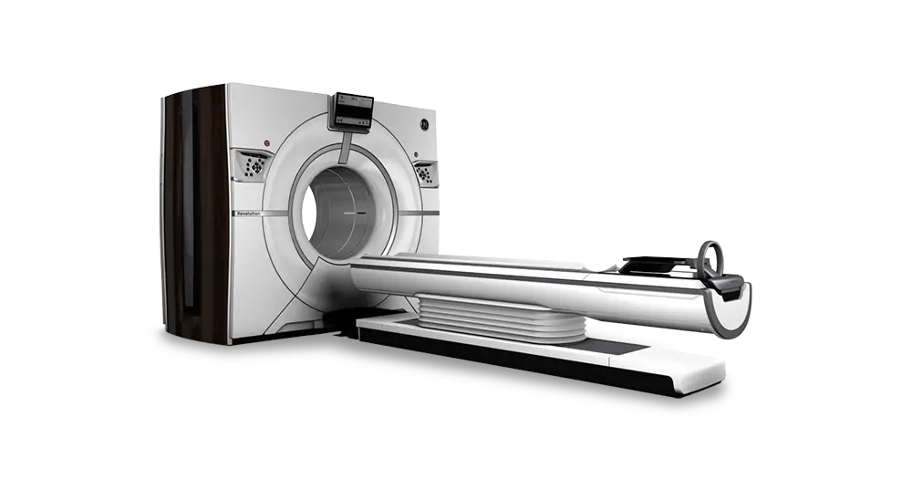
A cardiac CT scan, better known as CT coronary angiography, is a medical imaging procedure that uses CT (Computed Tomography) scanning technology to produce three-dimensional images of the heart and coronary arteries.
The process involves injecting contrast dye into a blood vessel, which allows the doctor to see blood flow and detect blockages or abnormalities in the arteries that supply blood to the heart. With highly detailed images, a cardiac CT scan can help diagnose various heart diseases more accurately.
How a Cardiac CT Scan Differs From Other Heart Tests
A cardiac CT scan differs from other heart tests, such as an ECG (electrocardiogram) and an echocardiogram, in the way it measures and assesses heart health.
- Electrocardiogram (ECG): This test records the heart’s electrical activity and can detect heart rhythm problems, but it does not give a physical picture of the structure of the heart and blood vessels. An ECG is often used as a first step to assess whether there is a rhythm abnormality or a heart attack.
- Echocardiogram: An ultrasound test that uses sound waves to produce images of the heart. It is useful for evaluating the heart’s pumping function and detecting damage to the valves or heart muscle. However, an echocardiogram cannot provide in-depth detail about the coronary arteries the way a cardiac CT scan can.
A cardiac CT scan is usually recommended when the doctor suspects coronary heart disease or when the risk of heart disease needs to be assessed in more depth.
Types of Cardiac CT Scans
There are several types of cardiac CT scans that can be performed, depending on the diagnostic purpose:
- CT Coronary Angiography: This is the cardiac CT scan used to examine the blood vessels of the heart. The procedure helps detect blockages or narrowing of the coronary arteries.
- CT Calcium Scoring: This test is used to assess the amount of calcium deposited in the coronary arteries, which is an indicator of potential heart disease risk. A high calcium score is associated with a high cardiovascular risk.
- CT for aortic diagnosis: This type is used to evaluate the aorta, the large artery that carries blood from the heart to the rest of the body, to diagnose conditions such as aneurysm and aortic dissection.
Benefits of a Cardiac CT Scan
1. Detecting Coronary Heart Disease (CHD)
A cardiac CT scan is one of the diagnostic tools that can help detect coronary heart disease (CHD). The procedure produces detailed images of the coronary arteries, allowing the doctor to see any blockage or narrowing in these blood vessels. This narrowing can be caused by plaque buildup (atherosclerosis), which restricts blood flow to the heart muscle.
Early detection of these blockages is very important because it can help prevent a heart attack and other complications. A cardiac CT scan helps the doctor determine how severe the blockage is and plan the right treatment, whether through lifestyle changes, medication, or interventional procedures such as angioplasty or heart bypass surgery.
2. Evaluating Calcium Plaque in the Coronary Arteries (Calcium Scoring)
CT calcium scoring is a special technique within cardiac CT scanning that focuses on measuring the amount of calcium in the walls of the coronary arteries. This calcium is an indicator of atherosclerotic plaque. The more calcium detected, the more likely there is plaque buildup and the higher a person’s risk of a future cardiovascular event.
CT calcium scoring results help the doctor assess an individual’s risk of developing heart disease, even before symptoms appear. This information is very valuable for making decisions about prevention and risk management, including lifestyle changes and preventive medication.
3. Assessing Heart Damage After a Heart Attack
After a heart attack, a cardiac CT scan can be used to assess how much the heart muscle has been damaged. A heart attack occurs when blood flow to part of the heart muscle stops, causing heart muscle cells to die (myocardial infarction). A cardiac CT scan can identify damaged areas of the heart muscle, helping the doctor determine the severity of the damage and predict the patient’s prognosis. This information is important for planning appropriate care and rehabilitation, and for managing the risk of long-term complications.
4. Diagnosing Other Heart Conditions
Besides CHD, a cardiac CT scan can also be used to diagnose various other heart conditions, including:
- Aortic aneurysm: An abnormal enlargement of the aorta (the main blood vessel leaving the heart).
- Aortic dissection: A tear in the wall of the aorta.
- Heart tumors: Abnormal growths in the heart.
- Congenital heart defects: Abnormalities in the structure of the heart present from birth.
- Heart valve disease: Disorders of heart valve function.
With its ability to produce detailed 3D images, a cardiac CT scan provides important information for diagnosing various heart conditions, helping the doctor decide on the most appropriate treatment plan.
The Cardiac CT Scan Procedure
1. Steps of the Examination
The cardiac CT scan process generally includes several stages:
- Before the examination: The doctor will ask about your medical history, including drug allergies, especially to the contrast dye that will be used. You may be asked to fast for a few hours before the examination, depending on the type of CT scan being done. You may also be asked to temporarily stop taking certain medications. The medical staff will explain the procedure in detail and answer your questions.
- During the examination: You will lie on an examination table that moves into the CT scanner. An IV line will be placed to inject contrast dye into your vein. You may feel a slight cold or warm sensation when the contrast dye is injected. During the scan, you will need to hold your breath for a few moments so that the images are clearer. The CT scanner will take images of your heart from various angles. This process generally takes a few minutes.
- After the examination: Once the examination is finished, the IV line will be removed. You can return to your normal activities unless the doctor gives special instructions. The results will be analyzed by the doctor and explained to you over the next few days.
2. Duration of the Examination
The overall duration of a cardiac CT scan procedure, including preparation and scanning time, usually ranges from 30 minutes to 1 hour. However, the time needed can vary depending on the type of examination and the patient’s condition.
3. Pain or Discomfort During the Examination
Most patients report feeling no pain during a cardiac CT scan. Some patients may feel slightly uncomfortable because they have to lie still for several minutes, or may feel a slight warm or cold sensation when the contrast dye is injected. However, this discomfort is usually mild and tolerable.
4. Risks and Side Effects
Like all medical procedures, a cardiac CT scan carries potential risks and side effects, although they are relatively low. Some possible side effects include:
- Allergic reaction to contrast dye: Some patients may have an allergic reaction to the injected contrast dye, ranging from a mild skin rash to a serious anaphylactic reaction. That is why your allergy history will be asked about carefully before the procedure.
- Radiation exposure: CT scans use ionizing radiation, although the dose is low and generally safe. However, radiation exposure can slightly increase the risk of cancer in the long term. The doctor will weigh the benefits and risks before deciding to carry out this procedure.
- Other side effects: Other rare side effects include nausea, vomiting, dizziness, and pain at the injection site.
Risks and Side Effects of a Cardiac CT Scan
Although a cardiac CT scan is generally safe, like other medical procedures, it has potential risks and side effects that you should understand.
1. Allergic Reactions to Contrast Dye
The contrast dye used in a cardiac CT scan, which is usually iodine-based, can cause allergic reactions in some people. These reactions can vary, from mild ones such as a skin rash, itching, or slight swelling, to more serious reactions such as shortness of breath, a drop in blood pressure (shock), or life-threatening anaphylaxis.
That is why, before the procedure, the doctor will ask about your allergy history in detail. If you have a significant history of allergies, especially to iodine or to contrast dye in the past, the doctor may prescribe anti-allergy medication before the procedure or even consider other options for heart imaging. If an allergic reaction occurs during the procedure, immediate medical treatment will be given.
2. Radiation Exposure
During a cardiac CT scan, the patient is exposed to X-ray radiation to obtain clear images of the coronary arteries. However, this radiation exposure is considered low and is balanced by the benefit of an accurate diagnosis. As a precaution, the doctor and technician will make sure that the radiation dose used is the minimum needed to obtain optimal results.
This procedure is also not recommended for pregnant women without careful consideration, because radiation can pose a risk to the fetus. With a careful approach, the risks from radiation can be minimized without compromising the quality of the examination.
Modern technology in CT scanners also continues to be developed to reduce the radiation dose needed. Your doctor will always weigh the balance between the benefits and risks of radiation before deciding to carry out the procedure.
3. Other Side Effects
Besides allergic reactions and radiation exposure, some other rare side effects may include:
- Nausea and vomiting: Some patients may experience nausea or vomiting, especially after the contrast dye is given.
- Dizziness: Dizziness can occur as a reaction to the contrast dye or from having to lie still for a long time during the examination.
- Pain at the injection site: Mild pain or bruising at the IV injection site can also occur.
These side effects are generally mild and temporary. If you have side effects that are bothersome or do not improve, contact your doctor right away.
It is important to always discuss the risks and benefits of a cardiac CT scan thoroughly with your doctor before deciding to undergo this procedure. Your doctor will help you understand the risks and benefits related to your specific health condition.
Interpreting Cardiac CT Scan Results
After a cardiac CT scan, the next step is understanding the results. Interpreting these results is the job of an experienced cardiologist.
How Doctors Interpret Cardiac CT Scan Results
The cardiologist will analyze the three-dimensional (3D) images of the heart and coronary arteries produced by the CT scanner. They will carefully examine the structure of the heart and the blood flow, and look for abnormalities such as:
- Narrowing or blockage of the coronary arteries: The doctor will measure the degree of narrowing and determine how severely blood flow to the heart is obstructed.
- Calcium plaque in the coronary arteries: The amount and location of calcium plaque will be measured and assessed to determine the risk of coronary heart disease.
- Structural abnormalities of the heart: The doctor will check for abnormalities in the structure of the heart, such as its size and shape, and the condition of the heart valves.
- Other anomalies: The doctor will look for any other anomalies that may affect heart health.
What Normal and Abnormal Findings Mean
- Normal results: A normal cardiac CT scan shows that the coronary arteries have no significant narrowing or blockage, the heart structure is normal, and blood flows smoothly to the heart. This indicates that the heart is in good condition.
- Abnormal results: Abnormal results may show narrowing or blockage of the coronary arteries, calcium plaque, structural heart abnormalities, or other anomalies. The doctor will explain how severe the abnormality is and discuss its implications for the patient’s heart health. Abnormal results require appropriate follow-up and treatment.
Next Steps After the Examination
The next steps after a cardiac CT scan depend on the results.
- Normal results: If the results are normal, the doctor will give recommendations for keeping your heart healthy through a healthy lifestyle, including a balanced diet, regular exercise, and avoiding smoking. Follow-up tests may be recommended depending on your individual risk factors.
- Abnormal results: If the results are abnormal, the doctor will explain the findings in detail and discuss appropriate treatment options. Treatment options may include lifestyle changes, medication (for example, cholesterol-lowering drugs or blood thinners), or interventional procedures such as angioplasty (a procedure to open a blocked artery) or heart bypass surgery (a procedure to route blood around the blocked part of an artery).
It is important to discuss your cardiac CT scan results and treatment plan thoroughly with your doctor. Your doctor will give a clear explanation and answer all your questions to make sure you get the right care.
Cost of a Cardiac CT Scan
The cost of a cardiac CT scan can vary depending on various factors, including the location of the hospital or clinic, the type of CT scan performed, and the technology used. The exact cost depends on the patient’s condition and the type of examination needed. Some health facilities may offer more affordable package prices, especially when the scan is done together with other health checks. Information on heart checkup packages that include a CT scan is available on the MCU Packages page.
Besides the cost of the procedure itself, there may also be additional costs, such as doctor consultation fees, follow-up tests, or the use of contrast dye, which may be charged separately.
Before having a cardiac CT scan, it is advisable to check with your healthcare provider and ask clearly about all the costs that may arise. Many hospitals also offer payment or insurance options that can help ease the cost burden. By understanding the costs involved, you can make better decisions about heart care for yourself or your family.
Conclusion
A cardiac CT scan is a non-invasive imaging procedure that produces detailed 3D images of the heart and coronary blood vessels. The procedure is useful for detecting coronary heart disease, assessing the risk of heart disease, assessing heart damage after a heart attack, and diagnosing various other heart conditions. Although it is generally safe, there are potential risks such as allergic reactions to contrast dye and radiation exposure, although these risks are relatively low. The procedure includes preparation before the examination, a short scan, and a quick recovery stage.
The decision to have a cardiac CT scan should be discussed with your doctor. This test may be recommended if you have risk factors for coronary heart disease, have symptoms such as chest pain or shortness of breath, or if your doctor suspects a heart abnormality based on the results of other tests. Your doctor will consider your overall health, including your family history and other risk factors, to determine whether a cardiac CT scan is the right test for you. Do not delay consulting a doctor if you have concerns about your heart health.
Protect yourself and your family from heart disease! Heart disease is a serious threat that can be prevented. By maintaining a healthy lifestyle, including a balanced diet, regular exercise, avoiding smoking, controlling your weight, and managing stress, you can reduce your risk of heart disease. Regular health checkups, including consultations with a doctor and heart tests when needed, are also very important for early detection and for preventing serious complications.
Keep as a reminder
Checklist before Your Consultation
Tick what you have prepared.
0 of 6 items checked
Checks are kept only while this page is open.
Next step
Want to Check Your Heart Health?
A checkup package helps identify risk factors early. What each package includes and its price are explained up front.
The information on this page is for educational purposes and does not replace medical consultation, diagnosis, or treatment. For any health concerns, please consult a doctor directly.
Doctors Related to This Topic

dr. Tigor Yehezkiel, Sp.Rad
Radiology
Cardiovascular Diagnostic Center
Clinical focusDiagnostic Radiology · CT Scan
View Profile
dr. Ika Ruriana, Sp.Rad
Radiology
Cardiovascular Diagnostic Center
Clinical focusDiagnostic Radiology · Radiography (X-Ray)
View Profile
Dr. med. dr. Denio A. Ridjab, Sp.JP(K)
Interventional Cardiology
Coronary Intervention Procedures +2 centers
Clinical focusInterventional Cardiology · Coronary Artery Disease
View ProfileFrequently Asked Questions.
Here is a frequently asked question about the safety of a cardiac CT scan.
Is a Cardiac CT Scan Safe?
A cardiac CT scan is generally safe, but like other medical procedures, it has potential risks and side effects. The biggest risk is an allergic reaction to the injected contrast dye, which can range from mild (skin rash) to serious (shortness of breath). There is also radiation exposure, although the dose is low and the diagnostic benefit usually outweighs this risk. Your doctor will assess your individual risk before the procedure and take the necessary precautions, such as giving anti-allergy medication if needed.
Sources and Transparency.
The information on this page is for educational purposes and does not replace medical consultation, diagnosis, or treatment. For any health concerns, please consult a doctor directly.
If you find information that needs clarifying, let us know through our Contact Us page.