
Quick answer
What Is Peripheral Stent Placement?
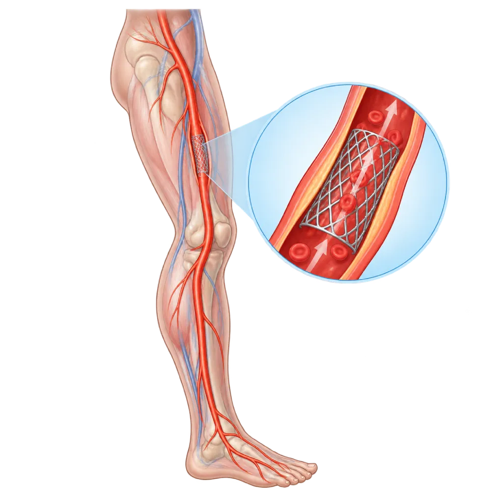
Peripheral stent placement is a catheter procedure to place a stent, a small metal mesh support, in a narrowed artery outside the heart, mainly the pelvic and leg arteries. The stent holds the artery wall open after balloon widening and stays inside the artery. The procedure takes about 1 to 2 hours under local anesthesia.
Sources01 / Understanding the procedure
About Peripheral Stent Placement
What Is Peripheral Stent Placement
A stent is a small metal mesh tube placed inside an artery to support its wall and keep it open. A peripheral stent is placed in an artery outside the heart, mainly the pelvic and leg arteries narrowed by plaque in peripheral artery disease. The stent is placed through a catheter, usually in the same procedure as balloon widening, which is covered on the Peripheral Angioplasty page. Once in place, the stent stays inside the artery and is gradually covered by a layer of cells from the vessel wall.
How It Works in Brief
The stent is carried in a folded state on the tip of a catheter to the narrowing. Some stents open on their own when their sheath is pulled back, while others are expanded with a balloon. The stent then presses the plaque against the wall and holds the artery wide open.
Types of Stents
- Self-expanding stents: made of flexible metal, suitable for the thigh artery, which moves and bends when you walk.
- Balloon-expandable stents: stiffer and can be positioned precisely, often used in the pelvic arteries and at the origin of the kidney arteries.
- Drug-eluting stents: release medication that slows the growth of scar tissue, reducing renarrowing.
- Covered stents: lined with a thin fabric-like material, used for example for complex blockages in the pelvic arteries or when the artery wall is torn.
When a Stent Is Placed
Not every narrowing needs a stent. A stent is usually placed if, after balloon widening, the artery wall tears, recoils, or is still narrowed, as well as for long narrowings or total blockages in the pelvic and thigh arteries. In the artery at the groin and behind the knee joint, stents are avoided whenever possible because these areas bend often. In the arteries below the knee, a balloon alone is used more often.
Stents in Other Arteries
Stents can also be placed in the arm arteries or kidney arteries. For kidney artery narrowing caused by plaque, large studies show that stents usually give no additional benefit compared with medication, so they are only considered in certain situations, for example when kidney function keeps getting worse or there are repeated episodes of sudden shortness of breath due to fluid in the lungs.
Living with a Stent
A stent cannot be felt from the outside and does not need to be replaced. Antiplatelet medications are taken to prevent clots inside the stent, and regular follow-up visits help detect renarrowing early. Checking the stent with ultrasound is covered on the Duplex Ultrasound page.
02 / Understanding the procedure
When Is This Procedure Considered?
A doctor may consider this procedure in the situations below. The decision is always made after a thorough assessment.
- Insufficient Balloon ResultThe artery wall tears, recoils, or is still narrowed after balloon widening.
- Narrowing or Blockage of the Pelvic ArteriesA stent is often placed right away because results last well in these large arteries.
- Long Narrowing in the Thigh ArteryA stent, often drug-eluting, helps the artery stay open longer.
- Tear or Leak in the Artery WallA covered stent can seal a tear or leak in the artery wall.
- Certain Narrowed Kidney or Arm ArteriesIf they cause symptoms or affect organ function, after evaluation by the team.
Situations to Consider First
- Narrowing in Areas That Bend OftenStents are avoided whenever possible in the artery at the groin and behind the knee joint.
- Unable to Take Antiplatelet MedicationThis medication is needed to prevent clots inside the stent.
- Good Result with a Balloon AloneA stent is not needed if the artery is already well opened with a balloon alone.
- Severe Allergy to Contrast DyeRequires special medication beforehand or a different treatment option.
- Active InfectionAn infection in the body or around the puncture site is treated first.
03 / Before the procedure
How Should You Prepare?
Peripheral stent placement is usually planned together with balloon widening, so the preparation is the same.
Blood vessel tests. An ABI test, duplex ultrasound, and CT angiography or angiography to map the arteries. These are covered on the Peripheral Angiography page.
Blood tests. Kidney function, complete blood count, and blood clotting.
Antiplatelet medications. Aspirin, and often clopidogrel, are started before the procedure, because both are important to prevent clots inside the stent. Tell the team if you have ever had bleeding problems or are taking other blood thinners.
Other medications and allergies. Tell the team about diabetes medications such as metformin, any contrast allergy, and any metal allergy.
Fasting. You usually do not eat for about 6 hours before the procedure. Drink water as instructed by the team.
Unsure about the preparation instructions? Ask our team before the procedure.
04 / During the procedure
How Is the Procedure Done?
The stent is placed in the catheterization lab under local anesthesia, and a mild sedative may be given. The procedure, including balloon widening, takes about 1 to 2 hours, longer for long or complex blockages.
Access and Mapping the Arteries
A small sheath is inserted into the artery in the groin, sometimes in the arm. Contrast dye is injected to see the location and length of the narrowing, and a blood thinner is given.
Widening with a Balloon
A guidewire is passed through the narrowing, then a balloon is inflated to prepare the site for the stent.
Placing the Stent
The folded stent is carried to the narrowing, its position is confirmed with X-rays, and then it is released or expanded with a balloon so it presses against the artery wall.
Checking the Result and Closing the Puncture
Angiography is repeated to confirm that the stent is fully open and blood flows smoothly down to the foot. The sheath is removed, then the puncture site is pressed or closed with a special device.
05 / Benefits and risks
Benefits and Risks Should Be Discussed Together.
Every medical procedure has benefits and risks. Your doctor will explain both for your situation before the procedure.
Benefits You Can Expect
- Keeps the Artery OpenThe stent supports the artery wall so it does not recoil or close again.
- Repairs a Torn Artery WallA tear after balloon widening is pressed back against the wall by the stent.
- No Large IncisionPlaced through a small puncture under local anesthesia, in the same procedure as the balloon.
- Less Renarrowing in Certain CasesEspecially for long narrowings in the thigh artery and with drug-eluting stents.
The benefits of a procedure are not a promise of a cure. Ask how the results will affect your care plan.
Risks You Should Know
- Renarrowing Inside the StentScar tissue can grow inside the stent; it is usually reopened with a balloon.
- Blood Clot in the StentRare, mainly if antiplatelet medication is stopped; it can block the stent suddenly.
- Bruising or Bleeding at the Puncture SiteUsually mild; rarely needs an additional procedure.
- Stent Fracture or MovementRare; more likely with stents in areas that bend often.
- Kidney Problems from Contrast DyeMore likely if kidney function is already reduced; prevented with IV fluids.
- Bleeding from Antiplatelet MedicationFor example, easy bruising or nosebleeds; the dose and duration are adjusted by your doctor.
Risks can differ from person to person. A full explanation and consent are given after an individual assessment.
06 / After the procedure
What Is Recovery Like?
Most patients stay one night for monitoring.
After the procedure. You lie down for a few hours with your leg straight, while the puncture site, foot pulses, and blood pressure are monitored.
Antiplatelet medications. Usually two types are taken for about 1 to 6 months, then one type long term, together with a statin. Do not stop them without consulting your doctor, because a clot can block the stent.
Activity. Light walking can start the next day. Avoid heavy lifting for about one week, then continue regular walking exercise.
Stent card. Keep the information about the type and location of your stent, and show it to staff before an MRI or other procedures.
Follow-up. An ABI test or duplex ultrasound is done regularly to monitor the stent.
Warning signs. Go to the emergency room right away if you have:
- Sudden pain, paleness, coldness, or numbness in the leg or foot
- Bleeding or growing swelling at the puncture site
- Fever, redness, or pus at the puncture site
- Black stools or bleeding that is hard to stop while taking antiplatelet medication
Know the Signs That Need Prompt Care.
Follow the team's guidance on warning signs. If severe or sudden symptoms appear after the procedure, go to the nearest emergency department right away or call Heart Attack Center 1500 258.
07 / Team and support
Doctors and Procedure Support
The doctors below are listed for this procedure. Listed by name, not by ranking.

dr. Emanoel Oepangat, Sp.JP(K)
Interventional Cardiology
Vascular Diagnostic and Therapeutic Center +1 center
Clinical focusCoronary Intervention · Peripheral Vascular Intervention
View Profile
dr. Suko Adiarto, Sp.JP(K), Ph.D.
Interventional Cardiology
Vascular Diagnostic and Therapeutic Center +1 center
Clinical focusCoronary Intervention · Peripheral Vascular Intervention
View ProfileVascular Interventional Suite
A procedure room with X-ray imaging for diagnosing and treating narrowed, blocked, or enlarged blood vessels outside the heart…
The availability of technology and test support is confirmed with the team. Technology is chosen according to clinical need.
08 / Common questions
Before You Plan the Procedure.
Frequently asked questions about peripheral stent placement.
What is the difference between a stent and balloon angioplasty?
Balloon angioplasty widens the artery, and then the balloon is removed. A stent is a metal mesh support that stays inside the artery to hold its wall open. Both are often done in a single procedure: the artery is first widened with a balloon, then a stent is placed if the balloon result is not sufficient or the location of the narrowing requires it. Balloon widening is covered on the Peripheral Angioplasty page.
Why doesn't every narrowing get a stent?
A stent does not always give a better result. In the artery at the groin and behind the knee joint, a stent can bend or break because these areas move often. In the arteries below the knee, a balloon alone is often enough. If a stent narrows again, treatment is also more complicated. For this reason, the doctor places a stent only when its benefits outweigh its drawbacks.
How long do I need to take antiplatelet medication?
Usually two antiplatelet medications, for example aspirin and clopidogrel, are taken for about 1 to 6 months, followed by one type long term. In some patients, the doctor may prescribe another combination based on the risk of clotting and bleeding. Do not stop the medication on your own, for example before a tooth extraction or surgery, without consulting the doctor who placed the stent.
Can a stent come loose, shift, or need replacing?
A stent attaches firmly to the artery wall and within a few weeks is covered by a layer of vessel cells, so it does not need to be replaced or removed. Shifting or breaking is rare and happens mainly when a stent is placed in an area that bends often. If renarrowing occurs inside the stent, it is usually reopened with a balloon through a catheter, including a drug-coated balloon.
Can I have an MRI after getting a stent?
The majority of peripheral stents allow MRI under certain conditions, but the requirements differ for each type. Keep the stent card or information from the hospital and show it to staff before an MRI, so the type of stent can be checked first. Also tell the team that you have a stent before any other procedure or surgery.
How long will I stay in the hospital, and when can I resume activities?
You usually stay one night. Light walking can start the next day, and daily activities usually resume within a few days. Avoid heavy lifting and strenuous exercise for about one week or as instructed by your doctor. Regular walking exercise, quitting smoking, and taking your medications as advised help keep the stent open.
Sources and Medical Review
Translated from the Indonesian edition medically reviewed by dr. Emanoel Oepangat, Sp.JP(K).
Sources as cited in the Indonesian edition.
- Mazzolai L, Teixido-Tura G, Lanzi S, et al. 2024 ESC Guidelines for the management of peripheral arterial and aortic diseases. Eur Heart J. 2024;45(36):3538-3700.
- Gornik HL, Aronow HD, Goodney PP, et al. 2024 ACC/AHA/AACVPR/APMA/ABC/SCAI/SVM/SVN/SVS/SIR/VESS Guideline for the Management of Lower Extremity Peripheral Artery Disease. Circulation. 2024;149(24):e1313-e1410.
- Schillinger M, Sabeti S, Loewe C, et al. Balloon angioplasty versus implantation of nitinol stents in the superficial femoral artery. N Engl J Med. 2006;354(18):1879-1888.
- Dake MD, Ansel GM, Jaff MR, et al. Durable clinical effectiveness with paclitaxel-eluting stents in the femoropopliteal artery: 5-year results of the Zilver PTX randomized trial. Circulation. 2016;133(15):1472-1483.
- Mwipatayi BP, Thomas S, Wong J, et al. A comparison of covered vs bare expandable stents for the treatment of aortoiliac occlusive disease. J Vasc Surg. 2011;54(6):1561-1570.
- Nordanstig J, James S, Andersson M, et al. Mortality with paclitaxel-coated devices in peripheral artery disease. N Engl J Med. 2020;383(26):2538-2546.
- Cooper CJ, Murphy TP, Cutlip DE, et al. Stenting and medical therapy for atherosclerotic renal-artery stenosis. N Engl J Med. 2014;370(1):13-22.